

Cone beam computed tomography - know its secrets
- PMID: 25859112
- PMCID: PMC4377156
Cone beam computed tomography - know its secrets
Abstract
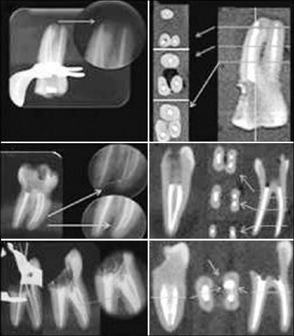
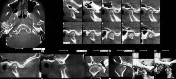
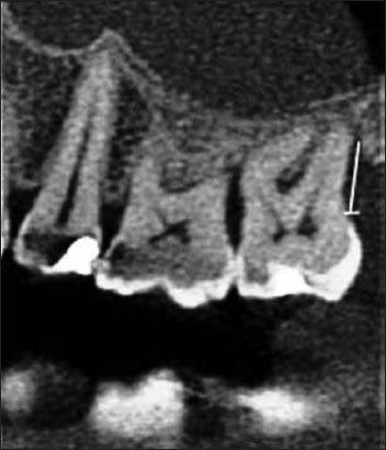
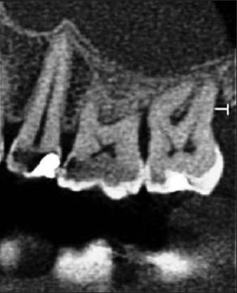
Cone-beam computed tomography (CBCT) is an advanced imaging modality that has high clinical applications in the field of dentistry. CBCT proved to be a successful investigative modality that has been used for dental and maxillofacial imaging. Radiation exposure dose from CBCT is 10 times less than from conventional CT scans during maxillofacial exposure. Furthermore, CBCT is highly accurate and can provide a three-dimensional volumetric data in axial, sagittal and coronal planes. This article describes the basic technique, difference in CBCT from CT and main clinical applications of CBCT.
Keywords: Cone-beam computed tomography; X-ray; diagnostic imaging; spiral computed tomography.
Conflict of interest statement
Figures
References
-
- Loubele M, Bogaerts R, Van Dijck E, Pauwels R, Vanheusden S, Suetens P, et al. Comparison between effective radiation dose of CBCT and MSCT scanners for dentomaxillofacial applications. Eur J Radiol. 2009;71(3):461–8. - PubMed
-
- Howerton WB, Jr, Mora MA. Use of conebeam computed tomography in dentistry. Gen Dent. 2007;55(1):54–7. - PubMed
-
- Winter AA, Pollack AS, Frommer HH, Koenig L. Cone beam volumetric tomography vs. medical CT scanners. N Y State Dent J. 2005;71(4):28–33. - PubMed
-
- Honda K, Larheim TA, Maruhashi K, Matsumoto K, Iwai K. Osseous abnormalities of the mandibular condyle: Diagnostic reliability of cone beam computed tomography compared with helical computed tomography based on an autopsy material. Dentomaxillofac Radiol. 2006;35(3):152–7. - PubMed
-
- Hashimoto K, Kawashima S, Araki M, Iwai K, Sawada K, Akiyama Y. Comparison of image performance between cone-beam computed tomography for dental use and four-row multidetector helical CT. J Oral Sci. 2006;48(1):27–34. - PubMed
Publication types
LinkOut - more resources
Full Text Sources