Review
. 2015 Mar;43(1):41-52.
doi: 10.2149/tmh.2014-25. Epub 2014 Nov 15.

The role of carbohydrates in infection strategies of enteric pathogens

Affiliations
Review

The role of carbohydrates in infection strategies of enteric pathogens

Kentaro Kato et al. Trop Med Health. 2015 Mar.

Abstract

Enteric pathogens cause considerable public health concerns worldwide including tropical regions. Here, we review the roles of carbohydrates in the infection strategies of various enteric pathogens including viruses, bacteria and protozoa, which infect the epithelial lining of the human and animal intestine. At host cell entry, enteric viruses, including norovirus, recognize mainly histo-blood group antigens. At the initial step of bacterial infections, carbohydrates also function as receptors for attachment. Here, we describe the function of carbohydrates in infection by Salmonella enterica and several bacterial species that produce a variety of fimbrial adhesions. During invasion by enteropathogenic protozoa, apicomplexan parasites utilize sialic acids or sulfated glycans. Carbohydrates serve as receptors for infection by these microbes; however, their usage of carbohydrates varies depending on the microbe. On the surface of the mucosal tissues of the gastrointestinal tract, various carbohydrate moieties are present and play a crucial role in infection, representing the site of infection or route of access for most microbes. During the infection and/or invasion process of the microbes, carbohydrates function as receptors for various microbes, but they can also function as a barrier to infection. One approach to develop effective prophylactic and therapeutic antimicrobial agents is to modify the drug structure. Another approach is to modify the mode of inhibition of infection depending on the individual pathogen by using and mimicking the interactions with carbohydrates. In addition, similarities in mode of infection may also be utilized. Our findings will be useful in the development of new drugs for the treatment of enteric pathogens.

Keywords: bacteria; carbohydrate; enteric pathogen; infection; protozoa; virus.


Figures

Fig. 1.
Fig. 1.
Schematic image of virus (in this case norovirus) recognition of carbohydrates on HBGAs at the host cell entry step. (A) Fucose (Fuc) and N-acetylgalactosamine (GalNAc) on A antigen are recognized by norovirus. (B) Fucose (Fuc) on O antigen is recognized by norovirus. (C) Fucose (Fuc) and galactose (Gal) on B antigen are recognized by norovirus. (D) Fucoses (Fuc) on Le b or y is recognized by norovirus.
Fig. 2.
Fig. 2.
General structure and localization of bacterial fimbriae and flagella. Gram-negative bacteria have many kinds of fimbriae (pili) and flagella. Bacterial lectin-like adhesive molecules (adhesins) included in bacterial fimbriae recognize and bind to sugar-containing molecules on the host cell surface. Bacterial fimbriae and adhesins contribute to bacterial attachment, the initial step of bacterial infection. Each bacterial adhesin recognizes a specific structure of its target sugar molecule, and bacterial fimbriae also help to determine the specificity (species, tissue, or cell) of bacterial infection. Two types of representative fimbriae of Salmonella and assortative bacteria (Type 1 fimbriae and Type 4 fimbriae) are shown. Type 1 fimbriae are short and highly expressed entirely on the surface of bacteria. Type 4 fimbriae are thin and flexible, expressed at low levels, and are generally located at the polar part of bacteria. FimA and FimH are categorized as Type 1 fimbriae. Long-polar fimbriae (LPF), Plasmid-code fimbriae (PEF) and bundle-forming pili (BFP) are categorized as Type 4 fimbriae. Std fimbriae are categorized as π-fimbriae. Bacterial flagella are the moving apparatus of bacteria, but their components can also contribute to the binding to sugar-containing molecules.
Fig. 3.
Fig. 3.
Various fimbriae of Salmonella and assortative bacteria and their sugar-containing receptor molecules. Salmonella and assortative bacteria express a variety of fimbriae. The minor component of Type 1 fimbriae, FimH, is present at the tip of type 1 fimbriae, mediates binding to D-mannose-containing structures and enables bacteria to colonize various host tissues [18]. Type 1 fimbria is highly expressed on the bacterial surface, allowing large amounts of bacteria to adhere via the FimH-mannose interaction. The various kinds of type 4 fimbriae play an important role in bacterial infection. Plasmid-encoded fimbria (PEF) is required for bacterial attachment to intestinal epithelial cells. PEF specifically binds to trisaccharide Galβ1-4(Fucα1-3)GlcNAc, also known as the Lewis X (LeX) blood group antigen [13]. Long polar fimbria (LPF) mediates the adhesion of S. typhimurium to murine Peyer’s patches [21]. Extracellular matrix proteins (ECMs) may act as receptors for LPF. ECMs are modified with various carbohydrate moieties, and the presence of Mannose inhibits the LPF-ECM interaction. Mannose-containing carbohydrates may participate in bacterial adhesion via LPF [23]. Std fimbriae are categorized as π-fimbriae and are well conserved among S. enterica serotypes but absent from other related bacterial species. Std fimbriae recognize and bind the H type 2 histo-blood group oligosaccharide, the terminal Fucα1-2Galβ1 moiety. S. typhi and S. paratifi can survive within the macrophages after they are engulfed by phagocytosis. Non-typhoidal Salmonella, S. typhimurium and S. enteritidis, however, are unable to survive within macrophages.
Fig. 4.
Fig. 4.
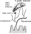
Schematic image of enteric pathogen (in this case T. gondii) invasion showing secreted proteins attached to carbohydrates on host cells. Some of the proteins from T. gondii tachyzoite (e.g., MICs, SAGs, P104) can be secreted or membrane-bound and attached to sialic acids, carbohydrates and glycoproteins during invasion.

References

    1. Spear PG, Shieh MT, Herold BC, et al. . Heparan sulfate glycosaminoglycans as primary cell surface receptors for herpes simplex virus. Adv Exp Med Biol 1992; 313: 341–353. - PubMed
    1. Ledeboer NA, Frye JG, McClelland M, et al. . Salmonella enterica serovar Typhimurium requires the Lpf, Pef, and Tafi fimbriae for biofilm formation on HEp-2 tissue culture cells and chicken intestinal epithelium. Infect Immun 2006; 74: 3156–3169. - PMC - PubMed
    1. Kobayashi K, Takano R, Takemae H, et al. . Analyses of interactions between heparin and the apical surface proteins of Plasmodium falciparum. Sci Rep 2013; 3: 3178. - PMC - PubMed
    1. Hutson AM, Atmar RL, Estes MK. Norovirus disease: changing epidemiology and host susceptibility factors. Trends Microbiol 2004; 12: 279–287. - PMC - PubMed
    1. Lindesmith L, Moe C, Marionneau S, et al. . Human susceptibility and resistance to Norwalk virus infection. Nat Med 2003; 9: 548–553. - PubMed