

A central mesencephalic reticular formation projection to the supraoculomotor area in macaque monkeys
- PMID: 25859632
- PMCID: PMC4600431
- DOI: 10.1007/s00429-015-1039-2
A central mesencephalic reticular formation projection to the supraoculomotor area in macaque monkeys
Abstract
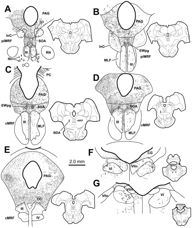
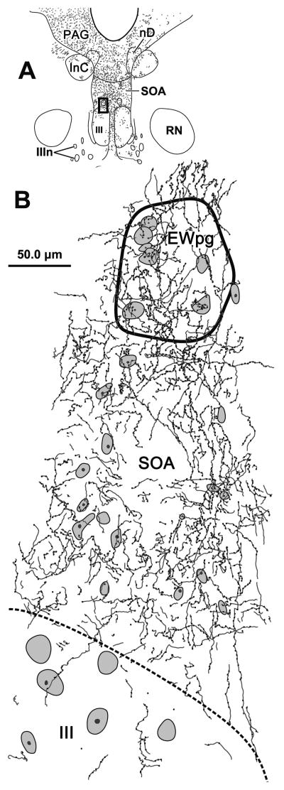
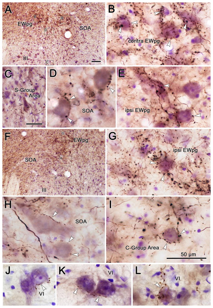
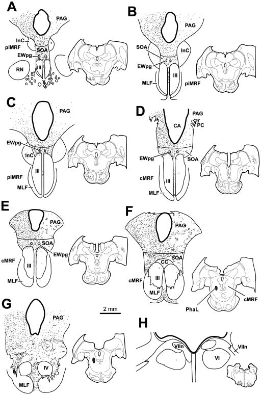
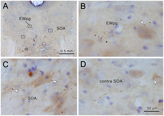
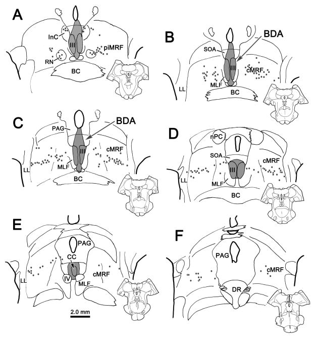
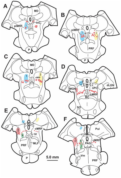
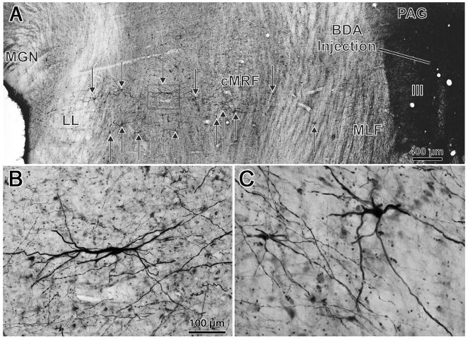
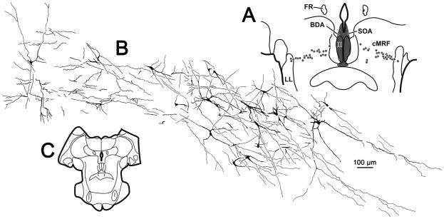
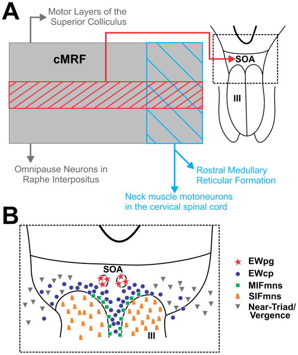
The central mesencephalic reticular formation is physiologically implicated in oculomotor function and anatomically interwoven with many parts of the oculomotor system's premotor circuitry. This study in Macaca fascicularis monkeys investigates the pattern of central mesencephalic reticular formation projections to the area in and around the extraocular motor nuclei, with special emphasis on the supraoculomotor area. It also examines the location of the cells responsible for this projection. Injections of biotinylated dextran amine were stereotaxically placed within the central mesencephalic reticular formation to anterogradely label axons and terminals. These revealed bilateral terminal fields in the supraoculomotor area. In addition, dense terminations were found in both the preganglionic Edinger-Westphal nuclei. The dense terminations just dorsal to the oculomotor nucleus overlap with the location of the C-group medial rectus motoneurons projecting to multiply innervated muscle fibers suggesting they may be targeted. Minor terminal fields were observed bilaterally within the borders of the oculomotor and abducens nuclei. Injections including the supraoculomotor area and oculomotor nucleus retrogradely labeled a tight band of neurons crossing the central third of the central mesencephalic reticular formation at all rostrocaudal levels, indicating a subregion of the nucleus provides this projection. Thus, these experiments reveal that a subregion of the central mesencephalic reticular formation may directly project to motoneurons in the oculomotor and abducens nuclei, as well as to preganglionic neurons controlling the tone of intraocular muscles. This pattern of projections suggests an as yet undetermined role in regulating the near triad.
Keywords: Edinger–Westphal; Eye movements; Midbrain; Oculomotor; Saccades; Vergence.
Conflict of interest statement
The authors declare that they have no conflicts of interest pursuant to the publication of this research.
Figures

Similar articles
-
A central mesencephalic reticular formation projection to medial rectus motoneurons supplying singly and multiply innervated extraocular muscle fibers.J Comp Neurol. 2017 Jun 1;525(8):2000-2018. doi: 10.1002/cne.24187. Epub 2017 Mar 14. J Comp Neurol. 2017. PMID: 28177529 Free PMC article.
-
A central mesencephalic reticular formation projection to the Edinger-Westphal nuclei.Brain Struct Funct. 2016 Nov;221(8):4073-4089. doi: 10.1007/s00429-015-1147-z. Epub 2015 Nov 28. Brain Struct Funct. 2016. PMID: 26615603 Free PMC article.
-
Internal organization of medial rectus and inferior rectus muscle neurons in the C group of the oculomotor nucleus in monkey.J Comp Neurol. 2015 Aug 15;523(12):1809-23. doi: 10.1002/cne.23760. Epub 2015 Apr 2. J Comp Neurol. 2015. PMID: 25684641 Free PMC article.
-
[Central oculomotor circuits].Rev Neurol (Paris). 1985;141(5):349-70. Rev Neurol (Paris). 1985. PMID: 3901182 Review. French.
-
Subcortical neural circuits for ocular accommodation and vergence in primates.Ophthalmic Physiol Opt. 1999 Mar;19(2):81-9. doi: 10.1046/j.1475-1313.1999.00434.x. Ophthalmic Physiol Opt. 1999. PMID: 10615444 Review.
Cited by
-
A central mesencephalic reticular formation projection to medial rectus motoneurons supplying singly and multiply innervated extraocular muscle fibers.J Comp Neurol. 2017 Jun 1;525(8):2000-2018. doi: 10.1002/cne.24187. Epub 2017 Mar 14. J Comp Neurol. 2017. PMID: 28177529 Free PMC article.
-
Transmitter inputs to different motoneuron subgroups in the oculomotor and trochlear nucleus in monkey.Front Neuroanat. 2015 Jul 24;9:95. doi: 10.3389/fnana.2015.00095. eCollection 2015. Front Neuroanat. 2015. PMID: 26257611 Free PMC article.
-
Response of supraoculomotor area neurons during combined saccade-vergence movements.J Neurophysiol. 2018 Feb 1;119(2):585-596. doi: 10.1152/jn.00193.2017. Epub 2017 Nov 15. J Neurophysiol. 2018. PMID: 29142092 Free PMC article.
-
Superior colliculus projections to target populations in the supraoculomotor area of the macaque monkey.Vis Neurosci. 2021;38:E017. doi: 10.1017/s095252382100016x. Epub 2021 Nov 11. Vis Neurosci. 2021. PMID: 36438664 Free PMC article.
-
An Anatomic Characterization of the Midbrain Near Response Neurons in the Macaque Monkey.Invest Ophthalmol Vis Sci. 2018 Mar 1;59(3):1486-1502. doi: 10.1167/iovs.17-23737. Invest Ophthalmol Vis Sci. 2018. PMID: 29625471 Free PMC article.
References
Publication types
MeSH terms
Grants and funding
LinkOut - more resources
Full Text Sources
Other Literature Sources
