

doi: 10.1038/srep09642.
The natural compound chebulagic acid inhibits vascular endothelial growth factor A mediated regulation of endothelial cell functions
Affiliations
- PMID: 25859636
- PMCID: PMC4819393
- DOI: 10.1038/srep09642
Item in Clipboard
The natural compound chebulagic acid inhibits vascular endothelial growth factor A mediated regulation of endothelial cell functions
Sci Rep.
.
Abstract
Vascular endothelial growth factor A (VEGFA) plays an important role in tumour angiogenesis and its angiogenic action is mainly mediated through its VEGF receptor 2 (VEGFR-2). Therefore drugs targeting VEGFA/VEGFR-2 are being presently used in the clinics for treatment of several types of solid malignant tumours. We here in report that low dose of chebulagic acid (CA), a hydrolysable tannin found in myrobalan fruits can inhibit VEGFA induced vascular permeability, endothelial cell proliferation, migration, tube formation and thereby, angiogenesis by suppressing VEGFR-2 phosphorylation. CA may thus be an effective and useful natural inhibitor of VEGFA mediated angiogenesis.
Figures
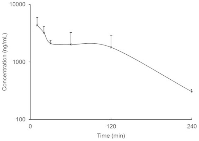
The plasma concentration-time profile of chebulagic acid in mice after oral ingestion of a single dose (100 mg/kg) of Triphala (n = 3). The error bars represent SEM.
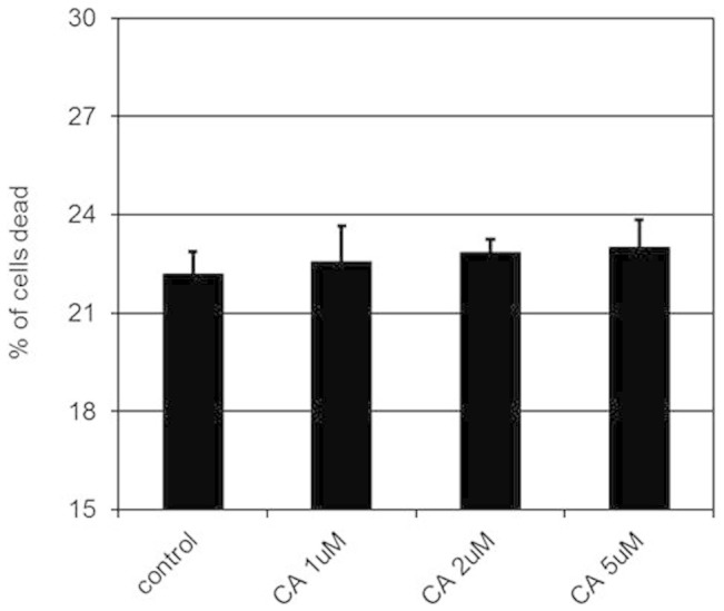
The cytotoxic effects of 1, 2 and 5 μM of chebulagic acid (CA) on human umbilical vein cells (HUVEC) (*, p < 0.05). The error bars represent SEM. The data represent six separate experiments.
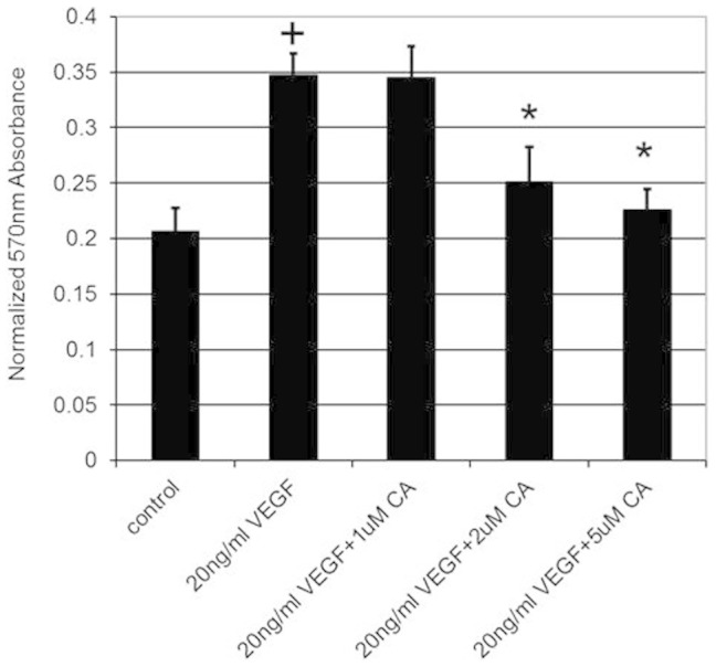
VEGFA stimulated proliferation of human umbilical vein cells (HUVEC) when compared to untreated control (+, p < 0.05). However, this stimulatory effect was inhibited when these cells were also treated with 2 and 5 μM of CA (*, p < 0.05). The error bars represent SEM. The data represent six separate experiments.
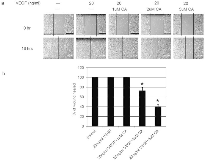
Phase-contrast photomicrographs demonstrated VEGFA induced complete wound closure or healing at 16 hr. On the contrary, this effect of VEGFA was abrogated when these cells were treated with 2 and 5 μM of CA. The wound healing was calculated as the distance covered by the cells in relation to the initial wound distance at 0 hr and is expressed as the percentage of initial distance at 0 hr. (*, P < 0.05). The error bars represent SEM. Scale bars in a, 200 um. The data represent six separate experiments.
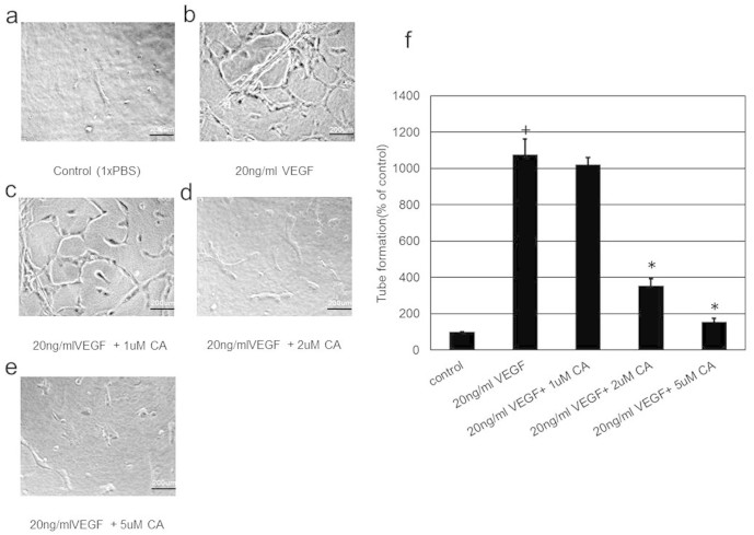
VEGFA stimulated tube formation in human umbilical vein cells (HUVEC) when compared to untreated control (+, p < 0.05). On the contrary, treatment with 2 and 5 μM of CA inhibited VEGFA induced endothelial cell tube formation (*, p < 0.05). Scale bars in (a), (b), (c), (d) and (e), 200 um. The data represent six separate experiments.
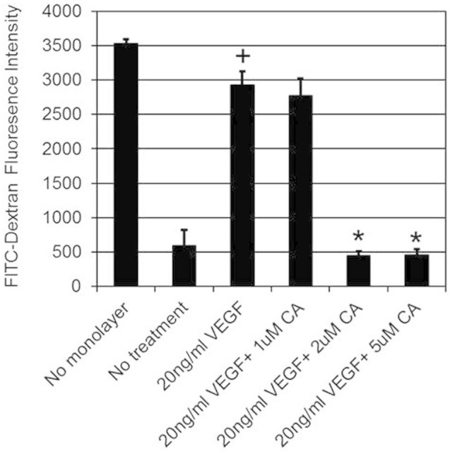
In comparison to untreated control, VEGFA induced significant permeability in human umbilical vein cells (HUVEC) (+, p < 0.05). On the contrary, treatment with 2 or 5 μM of CA significantly inhibited VEGFA stimulated permeability in HUVEC (*, p < 0.05). The data represent six separate experiments.
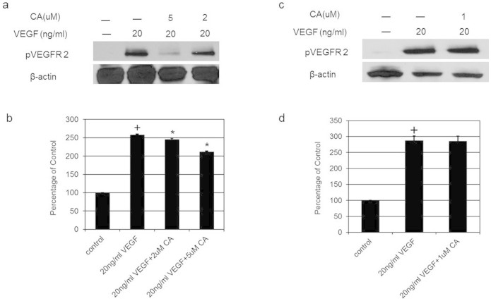
Western blot analysis shows that in comparison to untreated control, VEGFA induced significant VEGFR-2 phosphorylation (+, p < 0.05). However, there was also a significant inhibition of VEGFA induced phosphorylation of VEGFR2 after treatment with 2 or 5 μM of CA (*, p < 0.05). The blot was re-probed with an antibody to β actin for comparison of equal protein load. Cropped gel images have been used in this figure and the gels were run under the same experimental conditions. The data represent six separate experiments. Full-length blots are presented in Supplementary Figure S1.
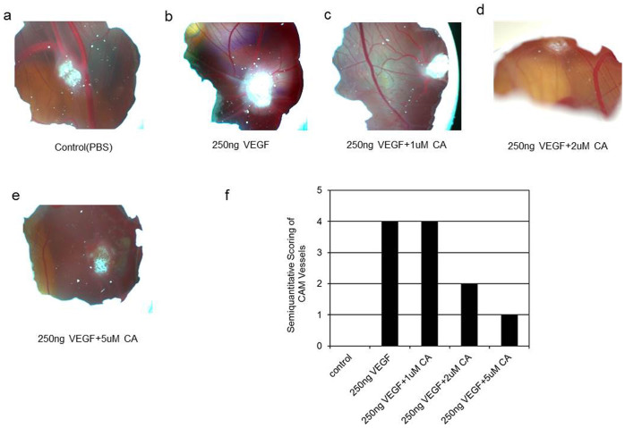
(a, f) Phosphate buffered saline (PBS) used as control did not induce blood vessel formation. (b, f) On the contrary, VEGFA stimulated new blood vessel formation. (c, f) 1 μM of CA did not inhibit VEGFA induced angiogenesis. (d, e, f) 2 or 5 μM of CA inhibited VEGFA induced new blood vessel formation. The photograph represents six separate experiments.
References
-
- Dvorak H. F. Vascular permeability factor/vascular endothelial growth factor: a critical cytokine in tumor angiogenesis and a potential target for diagnosis and therapy. J. Clin. Oncol. 20, 4368–4380 (2002). - PubMed
-
- Ferrara N. Vascular endothelial growth factor. Arterioscler. Thromb. Vasc. Biol. 29, 789–791 (2009). - PubMed
-
- Sloan B. & Scheinfeld N. S. Pazopanib, a VEGF receptor tyrosine kinase inhibitor for cancer therapy. Curr. Opin. Investig. Drugs. 9, 1324–1335 (2008). - PubMed
-
- Niraula S., Amir E., Vera-Badillo F., Seruga B., Ocana A. & Tannock I. F. Risk of incremental toxicities and associated costs of new anticancer drugs: a meta-analysis. J. Clin. Oncol. 32, 3634–3642 (2014). - PubMed
Publication types
MeSH terms
Substances
Grants and funding
LinkOut - more resources
Full Text Sources
Other Literature Sources
