. 2015 Apr 10;9(4):e0003663.
doi: 10.1371/journal.pntd.0003663. eCollection 2015 Apr.

Expression and function of S100A8/A9 (calprotectin) in human typhoid fever and the murine Salmonella model

Affiliations

Expression and function of S100A8/A9 (calprotectin) in human typhoid fever and the murine Salmonella model

Hanna K De Jong et al. PLoS Negl Trop Dis. .

Abstract

Background: Typhoid fever, caused by the Gram-negative bacterium Salmonella enterica serovar Typhi, is a major cause of community-acquired bacteremia and death worldwide. S100A8 (MRP8) and S100A9 (MRP14) form bioactive antimicrobial heterodimers (calprotectin) that can activate Toll-like receptor 4, promoting lethal, endotoxin-induced shock and multi-organ failure. We aimed to characterize the expression and function of S100A8/A9 in patients with typhoid fever and in a murine invasive Salmonella model.

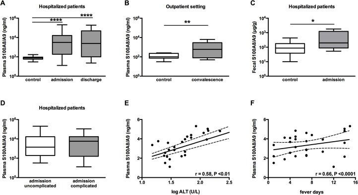
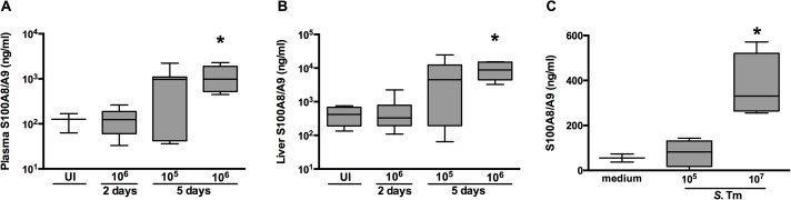
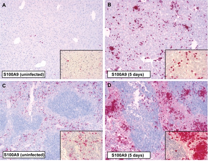
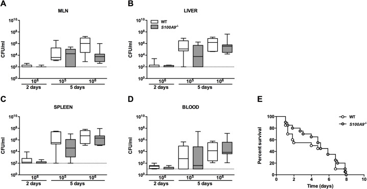
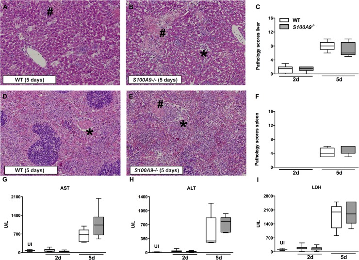
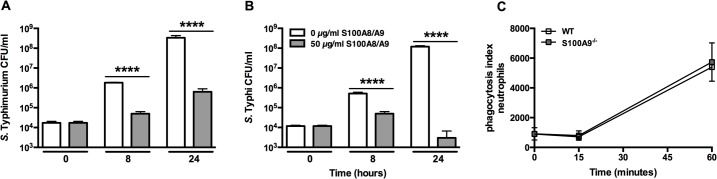
Methods and principal findings: S100A8/A9 protein levels were determined in acute phase plasma or feces from 28 Bangladeshi patients, and convalescent phase plasma from 60 Indonesian patients with blood culture or PCR-confirmed typhoid fever, and compared to 98 healthy control subjects. To functionally characterize the role of S100A8/A9, we challenged wildtype (WT) and S100A9-/- mice with S. Typhimurium and determined bacterial loads and inflammation 2- and 5- days post infection. We further assessed the antimicrobial function of recombinant S100A8/A9 on S. Typhimurium and S. Typhi replication in vitro. Typhoid fever patients demonstrated a marked increase of S100A8/A9 in acute phase plasma and feces and this increases correlated with duration of fever prior to admission. S100A8/A9 directly inhibited the growth of S. Typhimurium and S. Typhi in vitro in a dose and time dependent fashion. WT mice inoculated with S. Typhimurium showed increased levels of S100A8/A9 in both the liver and the systemic compartment but S100A9-/- mice were indistinguishable from WT mice with respect to bacterial growth, survival, and inflammatory responses, as determined by cytokine release, histopathology and organ injury.

Conclusion: S100A8/A9 is markedly elevated in human typhoid, correlates with duration of fever prior to admission and directly inhibits the growth of S. Typhimurium and S. Typhi in vitro. Despite elevated levels in the murine invasive Salmonella model, S100A8/A9 does not contribute to an effective host response against S. Typhimurium in mice.


Conflict of interest statement

The authors have declared that no competing interests exist.

Figures

Fig 1
Fig 1. S100A8/A9 complexes are abundantly present in plasma and feces from patients with typhoid fever and correlate with duration of fever.
Increased levels of S100A8/A9 (ng/ml) complexes are measured in plasma of patients with confirmed typhoid fever caused by S. Typhi admitted to the hospital (A; n = 28 for admission) and compared to healthy controls (n = 38). However S100A8/A9 levels were not yet normalized at time of discharge (n = 15). In a community based control study during convalescence (3 weeks post infection) S100A8/A9 remains elevated (B; n = 60) compared to healthy controls (n = 60). Increased levels of S100A8/A9 (μg/g) measured in feces from hospitalized patients (C; n = 14) compared to healthy controls (n = 36). No differences were seen in levels of S100A8/A9 between hospitalized patients with complicated or uncomplicated typhoid fever as detailed in method section (D). Data are expressed as box-and-whisker diagrams depicting the smallest observation, lower quartile, median, upper quartile and largest observation. Correlation curve log alanine aminotransferase (ALT) versus log plasma S100A8/A9 (E). Correlation curve days of fever prior to admission versus log plasma S100A8/A9 (F). For linear regression analysis, P value and Spearman rho are reported.
Fig 2
Fig 2. S100A/A9 is up regulated in a murine Salmonella model.
Levels of S100A8/A9 complexes measured in plasma (A) and liver homogenates (B) from wildtype (WT) mice orally challenged with 1x10⁵ or 1x10⁶ CFU S. Typhimurium after 0 (uninfected; UI; n = 3 per group), 2 or 5 days of infection (n = 5–8 per group). (C) S100A8/A9 complexes measured in murine whole blood 4 hours after stimulation with 1x10⁵ or 1x10⁷ heat inactivated S. Typhimurium (ATCC) diluted in RPMI to a final volume of 200 μl (n = 4). Data are expressed as box-and-whisker diagrams depicting the smallest observation, lower quartile, median, upper quartile and largest observation. * P<0.05, determined using Kruskal-Wallis testing.
Fig 3
Fig 3. Immunostaining for S100A9 in the liver and spleen of mice during S. Typhimurium infection.
Positive immunostaining for S100A9 in liver parenchyma (A) and the red pulp of the spleen (C) tissue was observed in uninfected control animals. Five days after infection with S. Typhimurium (106) there was a marked increase of S100A9 in both liver (B) and spleen (D) corresponding with inflammatory cell influx. Magnification 10 × and 40 ×.
Fig 4
Fig 4. S100A9 -/- mice are equally susceptible to S. Typhimurium infection when compared to wildtype mice in a Salmonella mouse model.
Bacterial loads in wildtype (WT; white bars) mice compared with S100A9 -/- mice (grey bars; data represent 5–8 mice per group) from mesenterial lymph nodes (MLN; A), liver and spleen homogenates (B, C), and blood (D), 2 and 5 days post infection with oral 1x10⁵ or 1x10⁶ CFU of S. Typhimurium. Data are expressed as box-and-whisker diagrams (log scale) depicting the smallest observation, lower quartile, median, upper quartile and largest observation. Survival curve of WT and S100A9 -/- mice after oral inoculation with 1x10⁶ CFU of S. Typhimurium (E; n = 20 mice per group in each experiment).
Fig 5
Fig 5. No effect of S100A8/A9 deficiency on organ injury during S. Typhimurium infection.
Representative slides of liver (A, B) and spleen (D, E) hematoxylin and-eosin (HE) staining of wildtype (WT) and S100A9 -/- mice, 5 days post infection. Liver and spleen from both WT and S100A9 -/- mice displayed advanced inflammation with necrosis (#) and thrombosis (*). Magnification 10 ×. Total pathology score was determined at indicated time points in WT (white bars) and S100A9 -/- (grey bars) mice according to the scoring system described in the methods section (C, F). Aspartate aminotransferase (AST; G), alanine aminotransferase (ALT; H), and lactate dehydrogenase (LDH; I) were measured in plasma. Data are expressed as box-and-whisker diagrams depicting the smallest observation, lower quartile, median, upper quartile and largest observation (5–8 mice per group at each time point).
Fig 6
Fig 6. S100A8/A9 inhibits bacterial growth of S. Typhimurium and S. Typhi in vitro.
Growth of S. Typhimurium (14028; A) and S. Typhi (Vivotif, Crucell; B) was assessed for a maximum of 24 hours in the presence of recombinant S100A8/A9 (50 μg/ml; grey bars) or control (white bars). Bacterial growth was dose dependently inhibited by S100A8/A9. Growth arrested, CFSE-labelled heat-inactivated S. Typhimurium bacteria were incubated with peripheral blood neutrophils (C) from wildtype (WT; white) and S100A9 -/- mice (grey) (n = 4 per mouse strain) for 0, 15 and 60 minutes respectively after which phagocytosis was quantified (see Methods). Data are expressed, as mean ± SD. **** P<0.0001, determined via non-parametric t tests comparing different dosages per time point.

References

    1. Crump JA, Mintz ED (2010) Global trends in typhoid and paratyphoid Fever. Clin Infect Dis 50: 241–246. 10.1086/649541 - DOI - PMC - PubMed
    1. Feasey NA, Dougan G, Kingsley RA, Heyderman RS, Gordon MA (2012) Invasive non-typhoidal salmonella disease: an emerging and neglected tropical disease in Africa. Lancet 379: 2489–2499. 10.1016/S0140-6736(11)61752-2 - DOI - PMC - PubMed
    1. de Jong HK, Parry CM, van der Poll T, Wiersinga WJ (2012) Host-pathogen interaction in invasive salmonellosis. PLOS pathog 8: e1002933 10.1371/journal.ppat.1002933 - DOI - PMC - PubMed
    1. Parry CM, Hien TT, Dougan G, White NJ, Farrar JJ (2002) Typhoid fever. N Engl J Med 347: 1770–1782. - PubMed
    1. Kawai T, Akira S (2010) The role of pattern-recognition receptors in innate immunity: update on Toll-like receptors. Nat Immunol 11: 373–384. 10.1038/ni.1863 - DOI - PubMed

Publication types

LinkOut - more resources