. 2015 Apr 9;161(2):255-63.
doi: 10.1016/j.cell.2015.02.042.

Horizontal transmission of clonal cancer cells causes leukemia in soft-shell clams

Affiliations

Horizontal transmission of clonal cancer cells causes leukemia in soft-shell clams

Michael J Metzger et al. Cell. .

Abstract

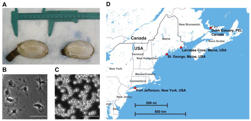
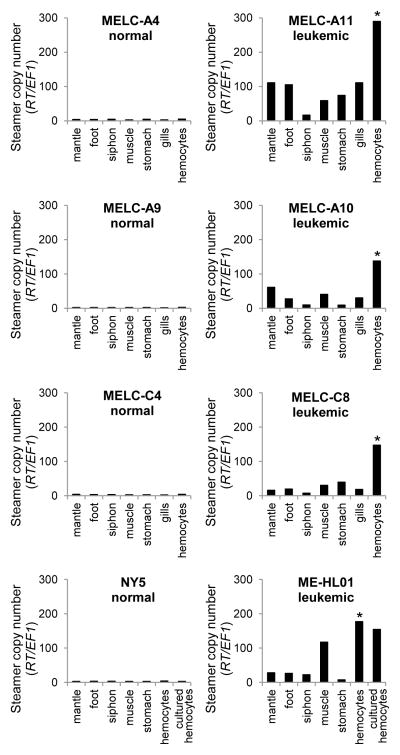
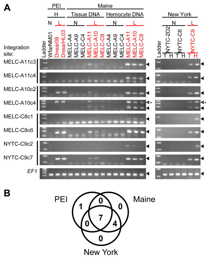
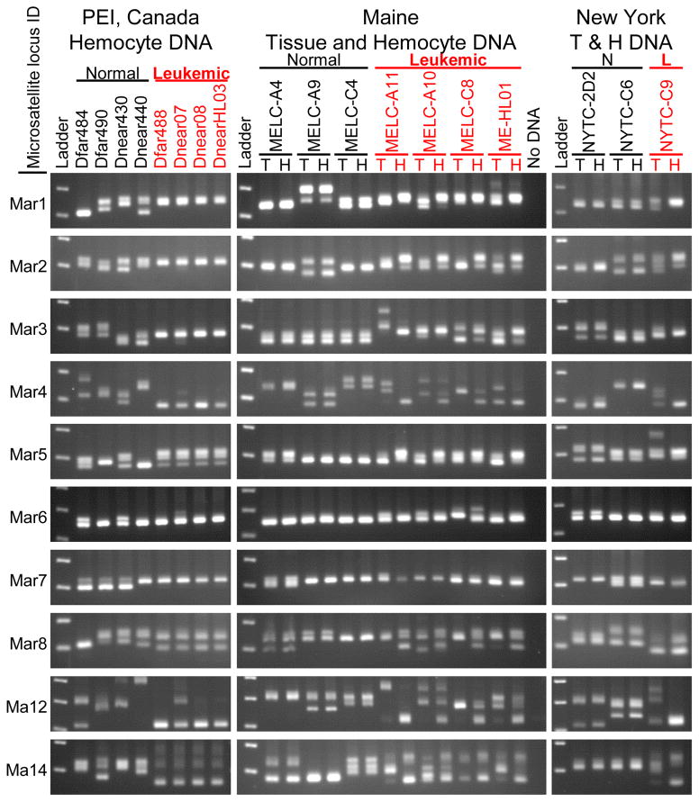
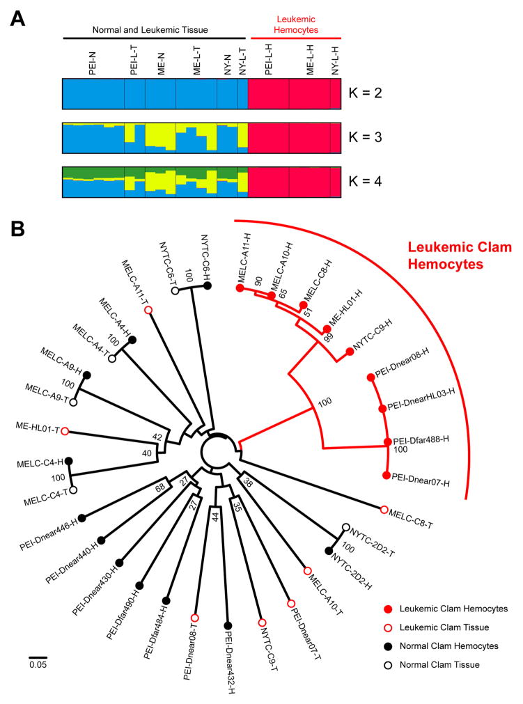
Outbreaks of fatal leukemia-like cancers of marine bivalves throughout the world have led to massive population loss. The cause of the disease is unknown. We recently identified a retrotransposon, Steamer, that is highly expressed and amplified to high copy number in neoplastic cells of soft-shell clams (Mya arenaria). Through analysis of Steamer integration sites, mitochondrial DNA single-nucleotide polymorphisms (SNPs), and polymorphic microsatellite alleles, we show that the genotypes of neoplastic cells do not match those of the host animal. Instead, neoplastic cells from dispersed locations in New York, Maine, and Prince Edward Island (PEI), Canada, all have nearly identical genotypes that differ from those of the host. These results indicate that the cancer is spreading between animals in the marine environment as a clonal transmissible cell derived from a single original clam. Our findings suggest that horizontal transmission of cancer cells is more widespread in nature than previously supposed.


Figures

Figure 1
Figure 1. Collection of normal and leukemic soft-shell clams (M. arenaria)
(A) Photograph of representative soft-shell clams (M. arenaria) with siphon partially extended. (B) Hemolymph from a normal clam (NYTC-C6), showing attachment of hemocytes to the dish and extension of pseudopodia. Scale bar: 50 μm. (C) Hemolymph from a heavily leukemic clam (NYTC-C9) showing lack of attachment and rounded, refractile morphology. Hemolymph of the leukemic clam was diluted 1:100 to allow visualization of single cells. Scale bar: 50 μm. (D) Map of the eastern coast of North America with the locations of the clam collection sites (Made with Mapbox Studio using data from OpenStreetMaps).
Figure 2
Figure 2. Steamer retrotransposon DNA copy numbers in different clam tissues
Quantitative PCR was used to determine the copy number of the Steamer retrotransposon in genomic DNA from hemocytes and solid tissues of four representative normal (left) and four leukemic (right) clams. Steamer copy number was quantitated using primers that amplify a region in the Steamer reverse transcriptase (RT) and was normalized to the single-copy gene EF1 (Siah et al., 2011). * indicates P < 0.01 for comparisons between neoplastic hemocytes and each other tissue tested (n = 3 for gills, n = 4 for other tissues), using two-tailed paired T-test of normalized Steamer copy number.
Figure 3
Figure 3. Clonal Steamer integration sites in neoplastic cells
(A) To determine the presence of specific integration sites in different animals, inverse PCR products were cloned and sequenced from clams from three locations (two integration sites per animal). For each integration site, a reverse primer was designed to match the flanking genomic sequence. Diagnostic PCR using a common forward primer in the Steamer LTR and an integration site specific reverse primer was used to determine the presence of each specific integration site in hemocyte (H) and tissue (T) DNA as indicated, of normal (N) and leukemic (L) clams. Sizes of the amplified DNAs were analyzed by agarose gel electrophoresis. Amplification of EF1 is shown as a control. Filled triangles mark the position of migration of the amplicon. An open triangle marks an unexpected PCR product. This band is due to amplification of a second Steamer integration site present in neoplastic cells of all populations. (B) Venn diagram of the number of cancer-specific integration sites shared by neoplastic cells from the three locations (out of the 12 sites tested, including 4 sites cloned from PEI samples previously (Arriagada et al., 2014)). None of the 12 sites were present in any normal animal tested.
Figure 4
Figure 4. Amplification of microsatellite loci from tissue and hemocyte DNA from normal and leukemic clams
PCR products using primers flanking ten microsatellite loci in hemocyte DNA and tissue DNA were displayed by electrophoresis on 2.5 % agarose gels and visualized by staining. Different alleles are determined by the sizes of the amplicons, with one band observed for animals homozygous at a particular locus, and two or more for heterozygotes and polyploid neoplastic cells. Each row of gels represents amplification from a single microsatellite locus, as labelled on the left. Hemocyte DNA is shown for normal and leukemic clams from PEI, Canada; and both Tissue (T) and Hemocyte (H) DNA is shown for Maine and New York clams. These data show that the leukemic hemocyte microsatellite alleles are identical to each other and distinct from their host tissue.
Figure 5
Figure 5. Analysis of microsatellite alleles
(A) CLUMPAK display of STRUCTURE (Pritchard et al., 2000) analysis of ten microsatellite loci showing the major population clustering with varying number of clusters (K) from K = 2 to 4. Each vertical bar represents a normal clam genotype or either tissue or hemocyte genotypes from leukemic clams with the colors representing cluster identity. (B) Neighbor-Joining tree constructed with genetic distances based on ten microsatellite loci using Bruvo’s method for analysis of loci from individuals with variable ploidy (Bruvo et al., 2004) calculated using the poppr package with R (Kamvar et al., 2014). Bootstrap values above 25 are shown at nodes. The scale bar represents a genetic distance of 0.05 (where 0 represents completely identical genotypes and 1 represents no common alleles). Each sample is marked as Tissue (T, closed circle) or Hemocytes (H, open circle) of Normal (Black) or Leukemic (Red) clams. The leukemic genotypes all cluster together in two branches clearly apart from the normal genotypes. Allele sizes are listed in Table S2 and further information is available in Supplementary Experimental Procedures.

Comment in

References

    1. AboElkhair M, Iwamoto T, Clark KF, McKenna P, Siah A, Greenwood SJ, Berthe FC, Casey JW, Cepica A. Lack of detection of a putative retrovirus associated with haemic neoplasia in the soft shell clam Mya arenaria. J Invertebr Pathol. 2012;109:97–104. - PubMed
    1. Arriagada G, Metzger MJ, Muttray AF, Sherry J, Reinisch C, Street C, Lipkin WI, Goff SP. Activation of transcription and retrotransposition of a novel retroelement, Steamer, in neoplastic hemocytes of the mollusk Mya arenaria. Proc Natl Acad Sci U S A. 2014;111:14175–14180. - PMC - PubMed
    1. Barber BJ. Neoplastic diseases of commercially important marine bivalves. Aquatic Living Resources. 2004;17:449–466.
    1. Beal BF, Kraus MG. Interactive effects of initial size, stocking density, and type of predator deterrent netting on survival and growth of cultured juveniles of the soft-shell clam, Mya arenaria L., in eastern Maine. Aquaculture. 2002;208:81–111.
    1. Brown RS, Wolke RE, Saila SB, Brown CW. Prevalence of neoplasia in 10 New England populations of the soft-shell clam (Mya arenaria) Ann N Y Acad Sci. 1978;298:522–534. - PubMed

Publication types

LinkOut - more resources