. 2015 Apr 9;161(2):277-90.
doi: 10.1016/j.cell.2015.02.016.

Organ-level quorum sensing directs regeneration in hair stem cell populations

Affiliations

Organ-level quorum sensing directs regeneration in hair stem cell populations

Chih-Chiang Chen et al. Cell. .

Abstract

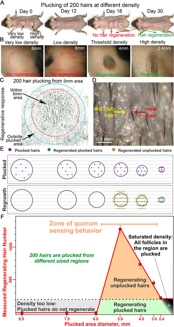
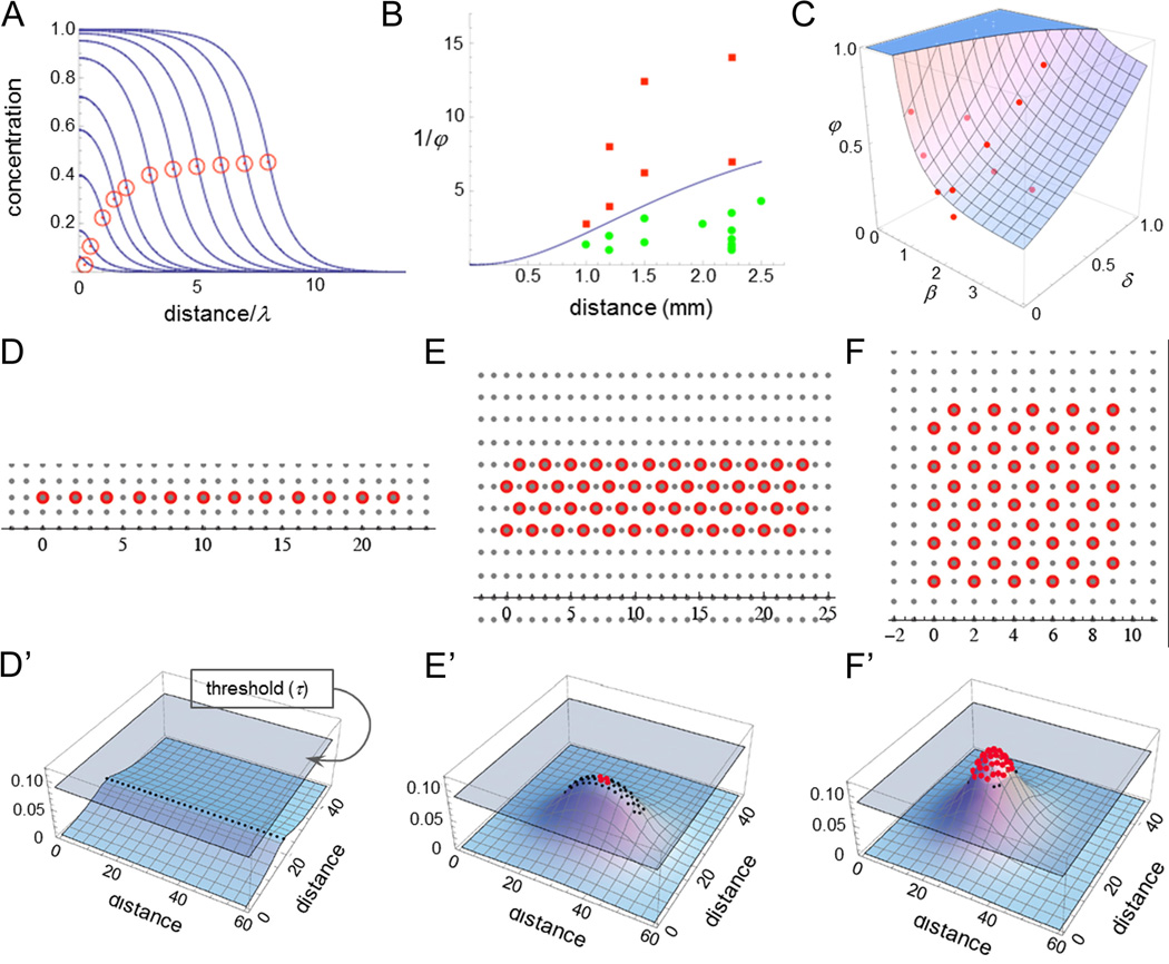
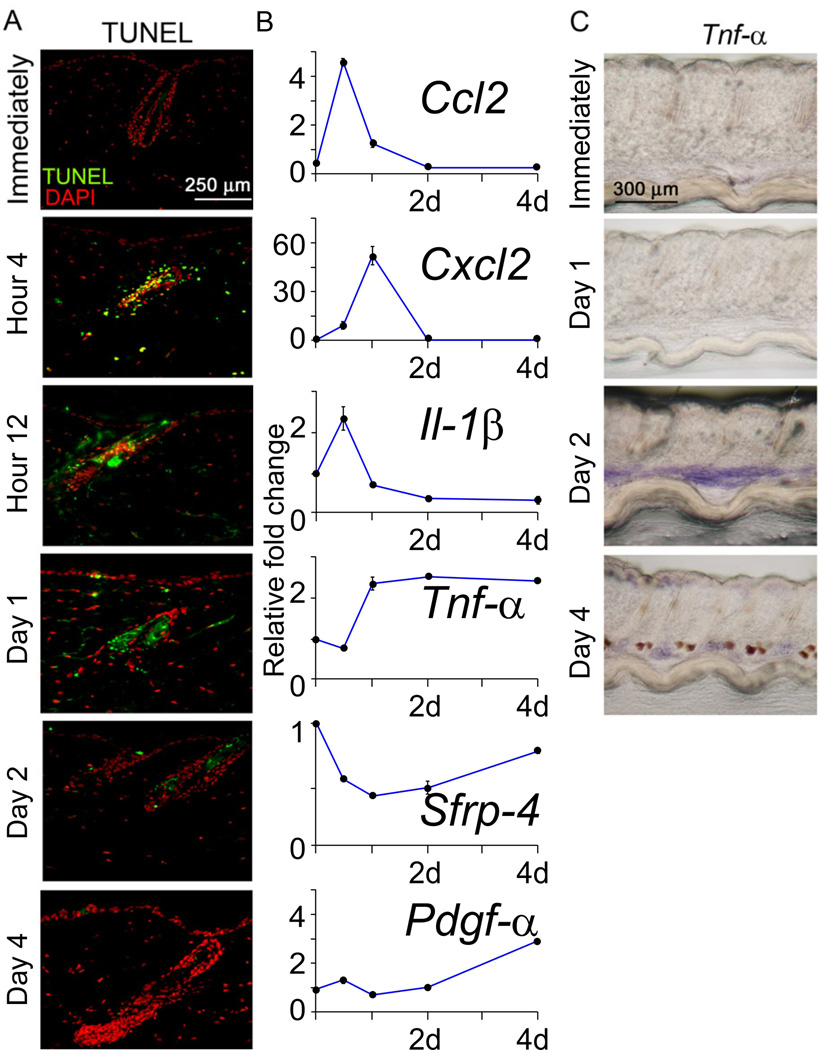
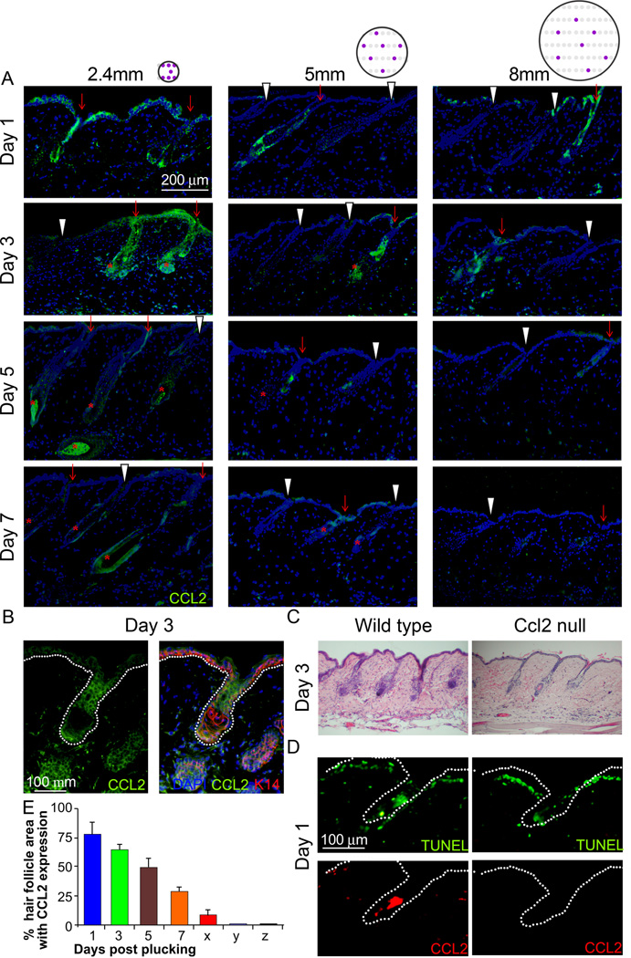
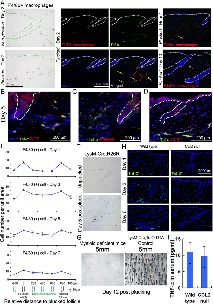
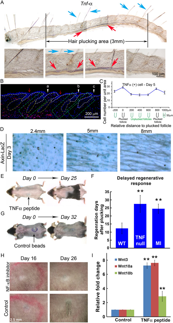
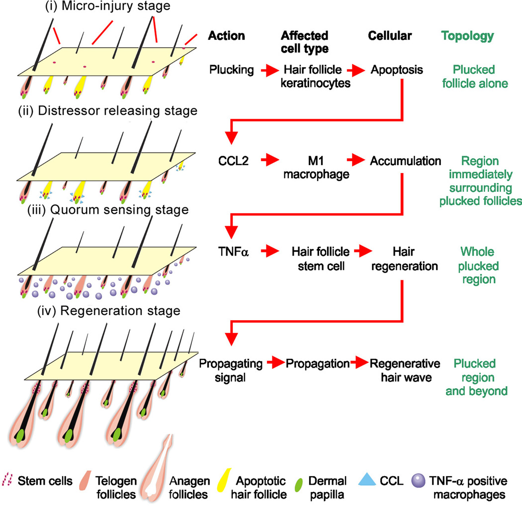
Coordinated organ behavior is crucial for an effective response to environmental stimuli. By studying regeneration of hair follicles in response to patterned hair plucking, we demonstrate that organ-level quorum sensing allows coordinated responses to skin injury. Plucking hair at different densities leads to a regeneration of up to five times more neighboring, unplucked resting hairs, indicating activation of a collective decision-making process. Through data modeling, the range of the quorum signal was estimated to be on the order of 1 mm, greater than expected for a diffusible molecular cue. Molecular and genetic analysis uncovered a two-step mechanism, where release of CCL2 from injured hairs leads to recruitment of TNF-α-secreting macrophages, which accumulate and signal to both plucked and unplucked follicles. By coupling immune response with regeneration, this mechanism allows skin to respond predictively to distress, disregarding mild injury, while meeting stronger injury with full-scale cooperative activation of stem cells.


Figures

Figure 1
Figure 1. Plucking-induced hair regeneration is a population based behavior which depends on the density and distribution of plucked-hair follicles within the unplucked follicle population
(A, B) Plucking 200 hairs from a circular 2.4 mm in diameter area (100% plucking) leads to hair regeneration 12 days later. Plucking 200 hairs in a 12 mm diameter area (100 mm2 area; low density plucking) fails to induce follicle regeneration even 30 days later. (C) Plucking induces regeneration of all follicles (the 200 plucked and 600 unplucked) within the plucked area (red circle, 5 mm in diameter). Unplucked follicles (400 HFs in total) outside the plucked area boundary then regenerate due to hair wave propagation (blue circle). (D) High power view showing unplucked follicle regeneration: the old gray club hair (yellow) is pushed out by the regenerating black anagen hair (red). (E) In this schematic drawing, grey dots represent telogen HFs. Black lines encircle exemplary plucked regions. Plucked follicles (purple dots). Regenerating plucked HFs (green dots). Regenerating unplucked HFs (tan dots). (F) Plot showing the hair regeneration response versus the size of the plucked field. For all different field sizes, 200 hairs are plucked evenly dispersed throughout the field. A regenerative response is observed when 200 hairs are plucked at a density above a threshold (10 hairs/mm2), which corresponds to plucking 200 hairs from a 5 mm diameter circular surface area (red line). Three responses represented by different colors (grey, tan, green), are observed (please see text for explanation). The quorum sensing zone is highlighted in orange. See also Figure S1, S2.
Fig. 2
Fig. 2. Mathematical modeling identifies the decay length of a putative quorum signal
(A) Calculated steady state concentrations for a diffusible substance produced within injury fields in proportion to the numbers of plucked HF. Each curve represents a different sized circular injury field, with the red circle placed at the value on the abscissa corresponding to the injury field radius, in units of the diffusing substance decay length. Specifically, the 11 curves represent increasing field sizes of 0.25, 0.5, 1, 1.5, 2, 3, 4, 5, 6, 7 and 8 decay lengths. As plucked regions grow larger, the value at the boundary asymptotes to one half the value at the center. λ is a decay length. Please see Results and supplements for more explanation. (B) Data from a variety of regeneration experiments involving circular wound fields are plotted as a function of the inverse of the plucked fraction (φ) and the radius of the wound field. The curve drawn between the points corresponding to cases of successful (green) and unsuccessful (red) regeneration was obtained from the equations that produced the curves in panel A, by fitting two parameters, the decay length and the threshold concentration for regeneration. The range of possible values consistent with the data was manually explored to yield a range of decay length estimates. (C) The same model was used as in panel B, but the data that were fit consisted of the distances, δ, just beyond the edges of injury fields at which initial regeneration was seen. (β, radius of the injury field; φ, the plucked fraction). The plotted surface represents a least-squares best fit to the data. (D–F, D’–F’) Effects of injury field shape. 50 hairs were plucked evenly, at a density of every other hair, either in a straight line (D, D’), a narrow rectangle (6:1 aspect ratio; E, E’) or a square (F, F’). In D’–F’, a discrete form of the equation used in panels A–C, in which each HF is modeled as a discrete source, was used to plot the steady-state spatial distributions of a distressor released by plucked follicles (distances are plotted in units of the inter-follicular distance, about 0.15 mm). Wherever plotted surfaces extend above a regeneration concentration threshold (grey plane), red dots mark the location of each HF indicating successful regeneration. The requirement that these curves be consistent with the observed regeneration patterns in all three cases was sufficient to provide yet a third estimate of the distressor decay length. See also supplement on mathematical model.
Figure 3
Figure 3. Identification of macro-environmental modulators following hair plucking
(A) TUNEL assay to measure apoptosis. (B) Real time PCR from extra-follicular macro-environmental tissues revealed the kinetics of gene expression induced by plucking (Normalized to GAPDH with 40 cycles, data are represented as mean +/− S.D., n=3). (C) Whole mount in situ hybridization showed that Tnf-α is markedly up-regulated in the inter-follicular area beginning 2 days after wax stripping. See also Figure S3, S4.
Figure 4
Figure 4. CCL2 is involved in plucking induced hair regeneration
(A) HF keratinocytes showed higher CCL2 expression (green) in plucked follicles (red arrow) than in unplucked follicles (white arrowhead). The circle with purple dots indicates the topology of plucked follicles (please also see Fig. 1E). Peak expression occurs 1–3 days after plucking, and no marked difference between the 2.4, 5 and 8mm groups were noted. (*: Regenerating HFs) (B) Double immunostaining for K14 and CCL2 of samples 3 days after plucking showed that HF keratinocytes in plucked follicles are the main source of CCL2. (C) Hair re-growth is retarded when hairs were plucked from CCL2 null mice. (D) CCL2 null mice showed similar apoptotic HF cells following plucking as wild type mice, but couldn’t induce CCL2 in apoptotic HF cells. (E) Graph showing the percentage of HF area expressing CCL2 at 1, 3, 5 and 7 days post plucking as well as unplucked HFs within (x) and outside (y) of the plucked field. CCL2 null mice do not express CCL2 (z). (n=3). (Data are represented as mean +/− S.D.). See also Figure S5.
Figure 5
Figure 5. CCL2 stimulates Tnf-α production by attracting CCR4 (+) M1 macrophages
(A) Tnf-α is up-regulated in the dermal macro-environment on day 2 after wax stripping. Tnf-α in the dermal macro-environment is produced by both dermal macrophages (F4/80+ cells, yellow arrow) and adipose cells (red arrow; Please see Fig. S5C). Few macrophages (yellow arrow) are present at hour 4 and day 10 after plucking. These macrophages do not express Tnf-α. (B, C) Staining shows that Tnf-α is mainly produced by M1 (iNOS positive) rather than M2 (Arginase positive) macrophages. (D) Tnf-α (+) cells express CCR4 in response to CCL2. (E) The number of F4/80+ cells is highest near plucked follicles and their density decreases with increasing distance from the plucked follicles. See Fig. S7A for the unit area we quantified for each data point. The number of F4/80+ cells is rapidly elevated at day 1 post-plucking, reaching a maximum at day 3 and then diminishing at 5 and 7 days after plucking. (F) LysM-Cre;R26R reporter mice show that the myeloid lineage-derived cells mostly are induced in the dermis around plucked HFs. (G) When 200 hairs were plucked from myeloid cell deficient mice from 5 mm region, hairs cannot be induced. (H) Tnf-α (+) cells was not induced in CCL2 null mice. (I) Tnf-α serum levels are similar between wild type and CCL2 null mice. (Data are represented as mean +/− S.D.). See also Figure S6–7.
Figure 6
Figure 6. Hair regeneration is proportional to the local concentration of Tnf-α
(A) Whole mount in situ hybridization shows Tnf-α (brown color in the dermis, red arrows) was induced under plucked and unplucked hairs (blue arrows) toward the center of the 3mm plucked zone 5 days after plucking. (B) Semi-quantitative assessment of the Tnf-α concentration using the 5mm group at day 5. Its expression level was quantified in 3 different skin regions (green boxes) that differ in their proximity to plucked follicles. “a” is closest to the plucked follicles and shows the highest Tnf-α levels. “b” is away from the plucked follicles and shows lower Tnf-α levels. “c” is furthest away and shows the least Tnf-α. (C) Quantitative assessment of the Tnf-α positive cells around plucked follicles and inter-plucked follicle dermis. See Fig. S7B for complete series. The pattern is similar to that of F4/80 macrophage distribution. (n=3) (D) Density-dependent plucking on Axin-LacZ mice show that the canonical Wnt/β-catenin signaling pathway was activated 3 days after plucking and the number of LacZ (+) HFs was proportional to the plucking density. (E) Subcutaneous injection of Tnf-α related peptide coated beads during refractory telogen can induce anagen re-entry and then propagate to the surrounding HFs. (F) Tnf-α null mice exhibit a 15-day delay in anagen re-entry following plucking of 200 hairs during refractory telogen phase. Intra-peritoneal macrophage inhibitor (MI) injection can also delay plucking induced hair regeneration by 12 days. (G) Albumin coated beads injection showed no anagen re-entry even after 32 days. (H) Subcutaneous NF-κB inhibitor injection can delay plucking-induced hair regeneration by 10 days. (I) Wnt3, Wnt10a and Wnt10b were activated in keratinocytes by TNF related peptides. (Data are represented as mean +/− S.D., ** p < 0.001). See also S6–7.
Fig. 7
Fig. 7. Molecular basis of quorum sensing behavior during the activation of hair stem cells in the follicle population
Schematic illustration of the process. Stage i: Minor-injury → hair keratinocyte apoptosis → CCL2 production. Stage ii: CCL2 secretion → macrophage accumulation, Stage iii: Macrophage and Tnf-α permeate the whole region. Stage iv: Tnf-α activates hair regeneration in the whole region. Hair regeneration further spreads due to propagation of regenerative hair waves. Please see text for more detail of the model.

Comment in

Similar articles

Cited by

References

    1. Ansell DM, Kloepper JE, Thomason HA, Paus R, Hardman MJ. Exploring the "hair growth-wound healing connection": anagen phase promotes wound re-epithelialization. J. Invest. Dermatol. 2011;131:518–528. - PubMed
    1. Bassler BL. Small talk. Cell-to-cell communication in bacteria. Cell. 2002;109:421–424. - PubMed
    1. Bischoff M, Gradilla AC, Seijo I, Andrés G, Rodríguez-Navas C, González-Méndez L, Guerrero I. Cytonemes are required for the establishment of a normal Hedgehog morphogen gradient in Drosophila epithelia. Nat. Cell Biol. 2013;15:1269–1281. - PMC - PubMed
    1. Cawthorn WP, Heyd F, Hegyi K, Sethi JK. Tumour necrosis factor-α inhibits adipogenesis via a beta-catenin/TCF4(TCF7L2)-dependent pathway. Cell Death. Differ. 2007;14:1361–1373. - PMC - PubMed
    1. Chen CC, Chuong CM. Multi-layered environmental regulation on the homeostasis of stem cells: the saga of hair growth and alopecia. J. Dermatol. Sci. 2012;66:3–11. - PMC - PubMed

Publication types

Associated data