. 2015 Apr 9;161(2):348-60.
doi: 10.1016/j.cell.2015.02.044.

Virulent Burkholderia species mimic host actin polymerases to drive actin-based motility

Affiliations

Virulent Burkholderia species mimic host actin polymerases to drive actin-based motility

Erin L Benanti et al. Cell. .

Abstract

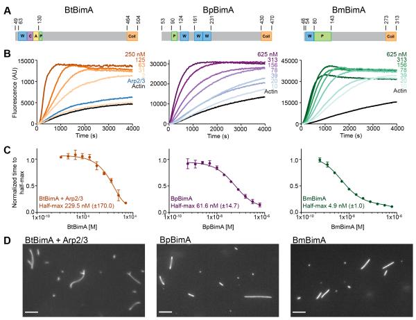
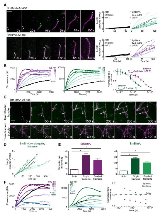
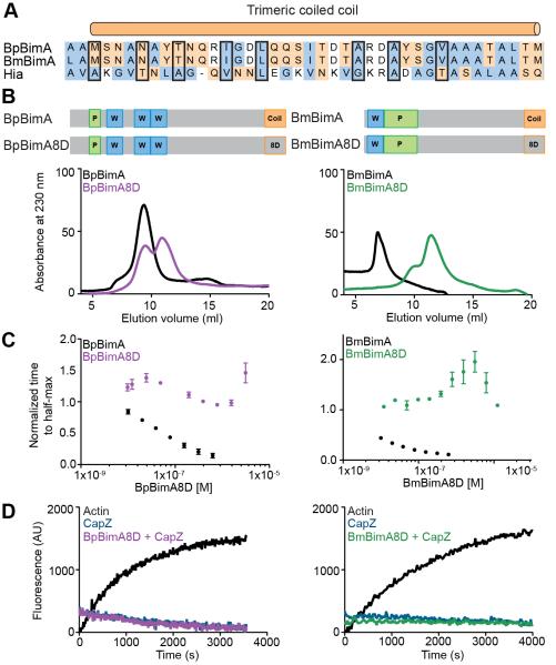
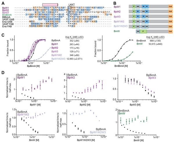
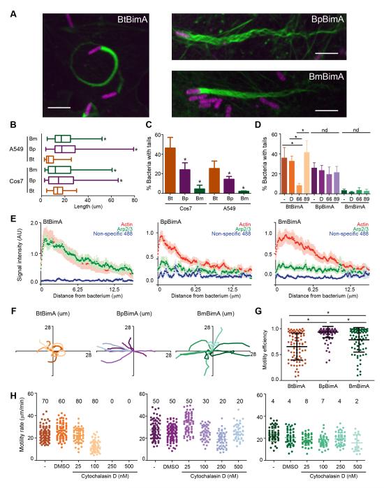
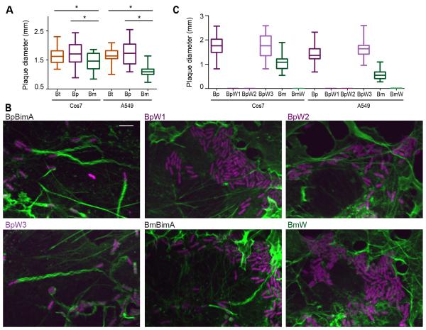
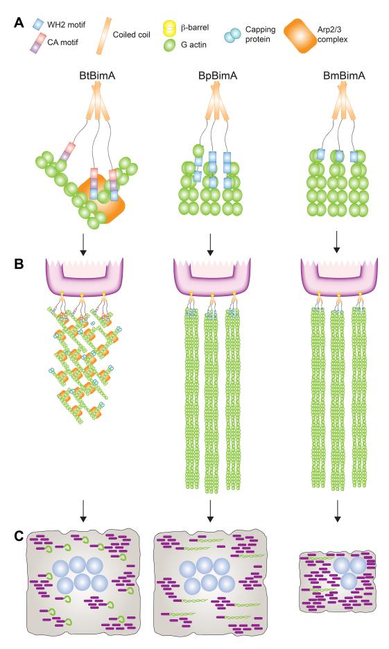
Burkholderia pseudomallei and B. mallei are bacterial pathogens that cause melioidosis and glanders, whereas their close relative B. thailandensis is non-pathogenic. All use the trimeric autotransporter BimA to facilitate actin-based motility, host cell fusion, and dissemination. Here, we show that BimA orthologs mimic different host actin-polymerizing proteins. B. thailandensis BimA activates the host Arp2/3 complex. In contrast, B. pseudomallei and B. mallei BimA mimic host Ena/VASP actin polymerases in their ability to nucleate, elongate, and bundle filaments by associating with barbed ends, as well as in their use of WH2 motifs and oligomerization for activity. Mechanistic differences among BimA orthologs resulted in distinct actin filament organization and motility parameters, which affected the efficiency of cell fusion during infection. Our results identify bacterial Ena/VASP mimics and reveal that pathogens imitate the full spectrum of host actin-polymerizing pathways, suggesting that mimicry of different polymerization mechanisms influences key parameters of infection.


Figures

Figure 1
Figure 1. BtBimA mimics host NPFs while BpBimA and BmBimA independently nucleate actin
(A) Domain schematics of BimA from different Burkholderia species. Actin-related and oligomerization sequences are shown. Numbers refer to amino acid position in full-length proteins. W, WASP homology 2; C, central; A, acidic; P, proline rich; Coil, trimeric coiled coil. (B) Pyrene actin polymerization reactions with increasing BimA concentrations with Arp2/3 complex (Bt) or in its absence (Bp, Bm). (C) The time to half-maximum fluorescence of the polymerization curves in (B) normalized to actin alone. The mean ± SD are shown and fits of each dataset are reported to estimate the concentration at which half-maximum activity is observed. (D) Epifluorescence images of polymerization reactions containing BtBimA with Arp2/3 complex, BpBimA or BmBimA after 15 min that were stabilized and stained with rhodamine phalloidin. Scale bars, 5 μm. See also Figure S1.
Figure 2
Figure 2. BmBimA and BpBimA processively bind growing filament barbed ends, increase elongation rates, bundle filaments and outcompete CapZ for barbed end binding
(A) Left, TIRF images showing BmBimA-AF488 and BpBimA-AF488 (green) and rhodamine-labeled actin (magenta). Time (s) is indicated. Scale bars, 3 μm. Right, filament length (number of subunits X 1000) over time for a minimum of 10 filaments with the mean elongation rate (sub/s ± SE) listed. (B) Left and middle, pyrene elongation assays with a range of BimA concentrations. Right, the time to half-maximum fluorescence normalized to actin alone for BmBimA (green) or BpBimA (purple) with the mean apparent KD ± SD listed. (C) TIRF images of BmBimA-AF488 (arrowhead) elongating two (top) or three (bottom) filaments (colors as in A). (D) Graph of filament length (number of subs X 1000) over time for pairs of co-elongating filaments (each pair is in matched colors of solid and dashed lines). (E) Mean elongation rates (sub/s ±SE) for actin alone or for single filaments or two- and three-filament bundles elongated with BmBimA-AF488 (left, green) or BpBimA-AF488 (right, purple). Asterisks denote paired samples that are significantly different (One-way ANOVA; P < 0.0001). (F) Left and middle, elongation reactions first incubated with 10 nM CapZ, followed by BimA and G-actin addition. Right, the time to half-maximum fluorescence normalized to actin alone (dashed black line) for BmBimA (green) or BpBimA (purple). See also Figure S2 and Movies S1-6.
Figure 3
Figure 3. BpBimA and BmBimA oligomerization is required for actin nucleation and barbed end binding
(A) Alignment of the trimeric coiled coils from BpBimA and BmBimA with the Hia trimeric coiled coil (Meng et al., 2006). Positions replaced with Asp residues in the BimA8D mutants are outlined in black. Hydrophobic residues are blue and charged residues orange. (B) Domain schematics of wild-type and BimA8D mutants in which eight positions were changed to Asp residues are shown above gel filtration elution profiles of wild-type and mutant proteins. (C) The time to half-maximum fluorescence normalized to actin alone in polymerization reactions with increasing BimA8D proteins. The means ± SD are shown with the wild-type data from Figure 1C for reference. (D) Elongation reactions with 10 nM CapZ and with our without BimA8D proteins.
Figure 4
Figure 4. WH2 requirements for BpBimA and BmBimA G-actin binding and nucleation
(A) Alignment of predicted WH2 sequences from BimA orthologs with known WH2 (GAB and FAB) sequences from human VASP and Drosophila Ena. The conserved α-helix and LKKT motif are indicated. Hydrophobic residues are blue and charged residues orange. Boxed residues were mutated to AA. (B) Domain schematics of WH2 mutant BimA proteins. Predicted WH2 motifs that were mutated are outlined in blue and denoted by AA. (C) Anisotropy measurements of monomeric actin488 binding to BimA. Data are represented by circles, and Hill equation fits are shown as solid lines. The means KD ± SD from at least two experiments are listed. (D) The time to half-maximum fluorescence normalized to actin alone in polymerization reactions with BimA. The means ± SD are shown with wild-type data from Figure 1C for reference.
Figure 5
Figure 5. BimA from different Burkholderia species mediates the formation of distinct actin tails and parameters of actin-based motility
(A) Merged images showing Cos7 cells infected with different Bt strains that constitutively express RFP (magenta). F-actin was stained with Alexa Fluor 488 phalloidin (green). Scale bars, 5 μm. (B) Mean actin tail lengths for the indicated strains in Cos7 and A549 cells. Boxes outline the 25th and 75th percentiles, midlines denote the medians and whiskers show minimum and maximum lengths. Asterisks denote lengths significantly different from BtBimA in that cell type (One-way ANOVA; P < 0.05). (C) Percent bacteria with actin tails (± SD). (D) Percent bacteria with actin tails (± SD) without or with 1 h treatment with DMSO, the Arp2/3 inhibitor CK-666 or the inactive compound CK-689. nd, no difference. (E) Mean actin, Arp2/3 or non-specific 488 nm fluorescence intensities (X 1000) from at least 10 actin tails (± SEM) are plotted along the first 12.5 μm of tail. (F) Tracks (ten per strain) depicting motility over 100 s for bacteria in Cos7 cells. (G) Motility efficiency from at least 80 tracks per strain, calculated as described in the text. (H) Motility velocity over 40 s for each strain in Cos7-Lifeact-EGFP cells untreated or treated with DMSO or CD. Mean frequencies of motile bacteria are listed. For all panels except (B), asterisks denote paired samples that are statistically different from one another (One-way ANOVA; P < 0.0001). See also Figure S3 and Movie S7.
Figure 6
Figure 6. BimA mechanisms of actin nucleation impact actin tail formation and host cell fusion
(A) Plaque diameters in Cos7 or A549 cell monolayers infected with Bt expressing wild-type BtBimA, BpBimA or BmBimA. (B) Merged images of Cos7 cells infected with wild-type or mutant BimA-expressing Bt strains that constitutively express RFP (magenta). F-actin was stained with Alexa Fluor 488 phalloidin (green). Scale bars, 5 μm. (C) Plaque diameters in Cos7 or A549 cell monolayers infected with Bt expressing wild-type or mutant BimA. (A, C) Boxes outline the 25th and 75th percentiles, midlines denote the medians and whiskers show minimum and maximum tail lengths. Asterisks denote significantly different sample pairs (One-way ANOVA; P < 0.05). See also Figure S4.
Figure 7
Figure 7. A model for BtBimA, BpBimA and BmBimA nucleation and actin tail and MNGC formation
(A) BtBimA activates host Arp2/3 complex using its WCA domain to nucleate branched actin networks. BpBimA nucleates and elongates filaments from barbed ends using its two N-terminal WH2 motifs, while BmBimA exhibits the same activities using its single WH2 motif to generate bundled filaments. (B) BtBimA and Arp2/3 form shorter tails with branched actin networks. BpBimA and BmBimA produce longer tails of bundled filaments. (C) Burkholderia synthesizing BtBimA or BpBimA form more actin tails and generate larger MNGCs compared with bacteria producing BmBimA.

Comment in

References

    1. Bachmann C, Fischer L, Walter U, Reinhard M. The EVH2 domain of the vasodilator-stimulated phosphoprotein mediates tetramerization, F-actin binding, and actin bundle formation. J. Biol. Chem. 1999;274:23549–23557. - PubMed
    1. Barzik M, Kotova TI, Higgs HN, Hazelwood L, Hanein D, Gertler FB, Schafer DA. Ena/VASP proteins enhance actin polymerization in the presence of barbed end capping proteins. J. Biol. Chem. 2005;280:28653–28662. - PMC - PubMed
    1. Bear JE, Svitkina TM, Krause M, Schafer DA, Loureiro JJ, Strasser GA, Maly IV, Chaga OY, Cooper JA, Borisy GG, et al. Antagonism between Ena/VASP proteins and actin filament capping regulates fibroblast motility. Cell. 2002;109:509–521. - PubMed
    1. Breitsprecher D, Kiesewetter AK, Linkner J, Urbanke C, Resch GP, Small JV, Faix J. Clustering of VASP actively drives processive, WH2 domain-mediated actin filament elongation. Embo J. 2008;27:2943–2954. - PMC - PubMed
    1. Breitsprecher D, Kiesewetter AK, Linkner J, Vinzenz M, Stradal TEB, Small JV, Curth U, Dickinson RB, Faix J. Molecular mechanism of Ena/VASP-mediated actin-filament elongation. Embo J. 2011;30:456–467. - PMC - PubMed

Publication types

MeSH terms