. 2015 Apr 30;6(12):10253-66.
doi: 10.18632/oncotarget.3520.

Endothelial exosomes contribute to the antitumor response during breast cancer neoadjuvant chemotherapy via microRNA transfer

Affiliations

Endothelial exosomes contribute to the antitumor response during breast cancer neoadjuvant chemotherapy via microRNA transfer

Nicolas Bovy et al. Oncotarget. .

Abstract

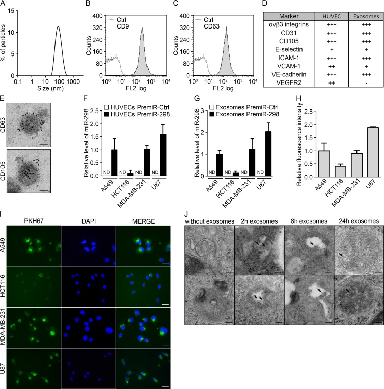
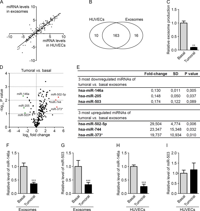
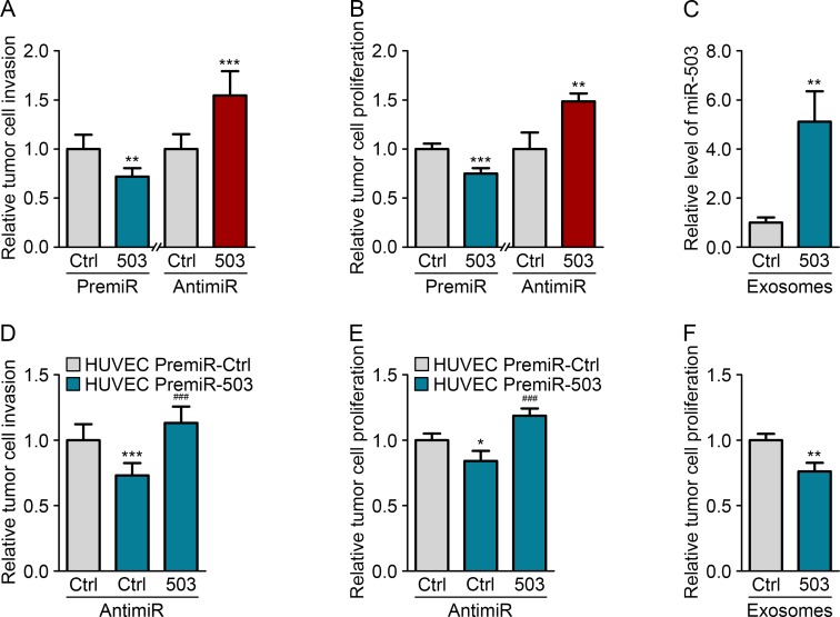
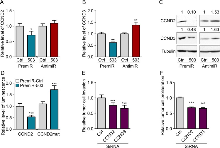
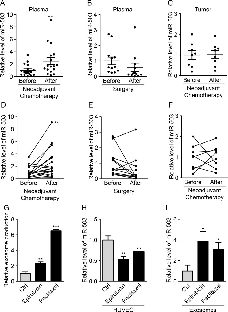
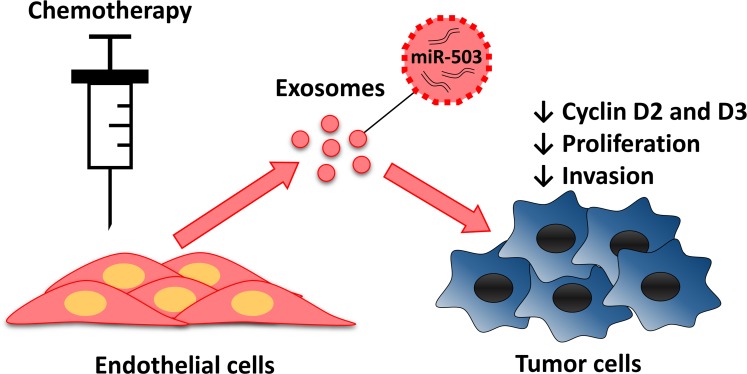
The interaction between tumor cells and their microenvironment is an essential aspect of tumor development. Therefore, understanding how this microenvironment communicates with tumor cells is crucial for the development of new anti-cancer therapies. MicroRNAs (miRNAs) are small non-coding RNAs that inhibit gene expression. They are secreted into the extracellular medium in vesicles called exosomes, which allow communication between cells via the transfer of their cargo. Consequently, we hypothesized that circulating endothelial miRNAs could be transferred to tumor cells and modify their phenotype. Using exogenous miRNA, we demonstrated that endothelial cells can transfer miRNA to tumor cells via exosomes. Using miRNA profiling, we identified miR-503, which exhibited downregulated levels in exosomes released from endothelial cells cultured under tumoral conditions. The modulation of miR-503 in breast cancer cells altered their proliferative and invasive capacities. We then identified two targets of miR-503, CCND2 and CCND3. Moreover, we measured increased plasmatic miR-503 in breast cancer patients after neoadjuvant chemotherapy, which could be partly due to increased miRNA secretion by endothelial cells. Taken together, our data are the first to reveal the involvement of the endothelium in the modulation of tumor development via the secretion of circulating miR-503 in response to chemotherapy treatment.

Keywords: angiogenesis; cancer; exosomes; miR-503; microRNAs.


Figures

Figure 1
Figure 1. Endothelial exosomes can transfer miRNAs to tumor cells
(A) Dynamic light scattering analysis of HUVEC exosomes (max = 94.93). Flow cytometry analysis of HUVEC exosomes immunolabeled for (B) CD9 and (C) CD63. (D) Table summarizing the levels of endothelial markers in HUVECs and HUVEC exosomes, measured using flow cytometry. (E) Electron micrographs of HUVEC exosomes labeled with CD63 and CD105, scale bars = 50 nm. (F) MiR-298 levels evaluated using qRT-PCR in cocultures either of tumor cells with HUVECs transfected with pre-miR-control or pre-miR-298 or (G) of tumor cells incubated with exosomes from HUVECs transfected with pre-miR-control or pre-miR-298. (H) Flow cytometry analysis of the uptake of exosomes (labeled with the green fluorescent PHK67 membrane linker) by tumor cells. (I) Fluorescence microscopy detection of the uptake of PHK67-labeled exosomes by tumor cells (DAPI, blue), scale bars = 25 μm. (J) Electron micrographs of MDA-MB-231 cell sections showing vesicles (arrows); after incubation with HUVEC exosomes for 0, 2, 8 and 24 hours, MDA-MB-231 cells showed larger multivesicular vesicles containing exosomes, scale bars = 100 nm. All data are the mean ± SD (n ≥ 3). *P < 0.05, **P < 0.01 and ***P < 0.001 vs. the respective control. Additionally, see Fig. S1.
Figure 2
Figure 2. The tumor environment modifies the export of a subset of endothelial miRNAs
(A) Dot plot of miRNA levels in HUVECs compared with HUVEC exosomes. (B) Diagram of miRNAs common and specific to HUVECs and HUVEC exosomes. (C) Exosome levels measured by protein quantification from the conditioned medium of HUVECs cultured in basal and tumor-mimicking medium conditions. (D) Volcano plot of fold changes (log2 values) and probability values (−log10) for individual miRNAs in the exosomes from HUVECs cultured under basal and tumoral conditions. (E) Table of the three most upregulated and downregulated miRNAs under tumoral vs. basal conditions. MiR-146a (F) and miR-503 (G) levels, evaluated by qRT-PCR in HUVEC exosomes cultured under tumoral or basal conditions. MiR-146a (H) and miR-503 (I) levels, evaluated by qRT-PCR in HUVECs cultured in tumoral or basal conditions. All data are the mean ± SD (A-E, n = 2; F-I, n = 3). *P < 0.05, **P < 0.01 and ***P < 0.001 vs. the respective control. Additionally, see Fig. S2.
Figure 3
Figure 3. Endothelial miR-503 impairs tumor growth in vitro
(A) Invasion level of MDA-MB-231 cells transfected with pre-miR-control or pre-miR-503 and with anti-miR-control or anti-miR-503. (B) Luminescence quantification of MDA-MB-231 cells transfected with pre-miR-control or pre-miR-503 and with anti-miR-control or anti-miR-503. (C) MiR-503 levels, measured by qRT-PCR in MDA-MB-231 cells incubated with 5 μg of HUVEC exosomes for 24 h. (D) Invasion level of MDA-MB-231 cells transfected with anti-miR-control or anti-miR-503 and cocultured with HUVECs transfected with pre-miR-control or pre-miR-503. (E) Luminescence quantification of MDA-MB-231 cells transfected with anti-miR-control or anti-miR-503 and cocultured with HUVECs transfected with pre-miR-control or pre-miR-503. (F) Luminescence quantification of MDA-MB-231 cells incubated with exosomes from HUVECs transfected with pre-miR-control or pre-miR-503. Additionally, see Fig. S3.
Figure 4
Figure 4. MiR-503 inhibits CCND2 and CCND3 expression of MDA-MB-231
Quantification of mRNA levels according to qRT-PCR of (A) CCND2 and (B) CCND3 in MDA-MB-231 cells transfected with pre-miR-503 or pre-miR-control and with anti-miR-control or anti-miR-503 and starved for 36 h. (C) Western blotting of CCND2 and CCND3 in MDA-MB-231 cells transfected with pre-miR-503 or pre-miR-control and with anti-miR-control or anti-miR-503 and starved for 36 h. (D) Luciferase activity from the CCND2 3′-UTR WT reporter plasmid and mutated NRAS 3′-UTR reporter plasmid cotransfected into MDA-MB-231 cells with pre-miR-control or pre-miR-503 after 24 hours. (E) Invasion levels of MDA-MB-231 cells transfected with siRNAs targeting CCND2 and CCND3. (F) Luminescence quantification of MDA-MB-231 cells transfected with siRNAs targeting CCND2 and CCND3. *P < 0.05, **P < 0.01 and ***P < 0.001 vs. the respective control. ###P < 0.001 vs. miR-503-exosomes with anti-miR-control. Additionally, see Fig. S4.
Figure 5
Figure 5. Neoadjuvant chemotherapy increases circulating miR-503 levels
(A) The miR-503 ratio according to qRT-PCR in blood samples of breast cancer patients before and after neoadjuvant chemotherapy. (B) The miR-503 ratio according to qRT-PCR in blood samples of breast cancer patients before and after surgery. (C) The miR-503 ratio according to qRT-PCR in tumor biopsies and residual tumors of breast cancer patients. (D) Individual follow-up of miR-503 levels, evaluated by qRT-PCR, in blood samples of breast cancer patients before and after neoadjuvant chemotherapy. (E) Individual follow-up of miR-503 levels, evaluated by qRT-PCR, in blood samples of breast cancer patients before and after surgery. (F) Individual follow-up of miR-503 levels, evaluated by qRT-PCR, in tumor biopsies and residual tumors of breast cancer patients. (G) Exosome levels, measured by protein quantification, from the conditioned medium of HUVECs treated with paclitaxel and epirubicin for 24 h. (H) The miR-503 levels, evaluated by qRT-PCR, in HUVECs treated with paclitaxel and epirubicin for 24 h. (I) The miR-503 levels, evaluated by qRT-PCR, in exosomes from HUVECs treated with paclitaxel and epirubicin for 24 h. *P < 0.05, **P < 0.01 and ***P < 0.001 vs. the respective control.
Figure 6
Figure 6. Endothelial transfer of miR-503 elicit antitumor response during neoadjuvant chemotherapy
Endothelial cells treated with chemotherapeutic agents release more exosomes that contain more miR-503. MiR-503 loaded exosomes induce a reduction of breast cancer cells proliferation and invasion caused by the inhibition of CCND2 and CCND3.

References

    1. Suzuki HI, Katsura A, Matsuyama H, Miyazono K. MicroRNA regulons in tumor microenvironment. Oncogene. 2014 - PMC - PubMed
    1. Goubran HA, Kotb RR, Stakiw J, Emara ME, Burnouf T. Regulation of tumor growth and metastasis: the role of tumor microenvironment. Cancer Growth Metastasis. 2014;7:9–18. - PMC - PubMed
    1. Carmeliet P. Angiogenesis in life, disease and medicine. Nature. 2005;438:932–936. - PubMed
    1. Ebos JM, Kerbel RS. Antiangiogenic therapy: impact on invasion, disease progression, and metastasis. Nat Rev Clin Oncol. 2011;8:210–221. - PMC - PubMed
    1. Li M, Li J, Ding X, He M, Cheng SY. microRNA and cancer. AAPS J. 2010;12:309–317. - PMC - PubMed

Publication types