. 2015:2015:460340.
doi: 10.1155/2015/460340. Epub 2015 Mar 10.

Intraoperative computed tomography versus Perdriolle and scoliometer evaluation of spine rotation in adolescent idiopathic scoliosis

Affiliations

Intraoperative computed tomography versus Perdriolle and scoliometer evaluation of spine rotation in adolescent idiopathic scoliosis

Rafal Pankowski et al. Biomed Res Int. 2015.

Abstract

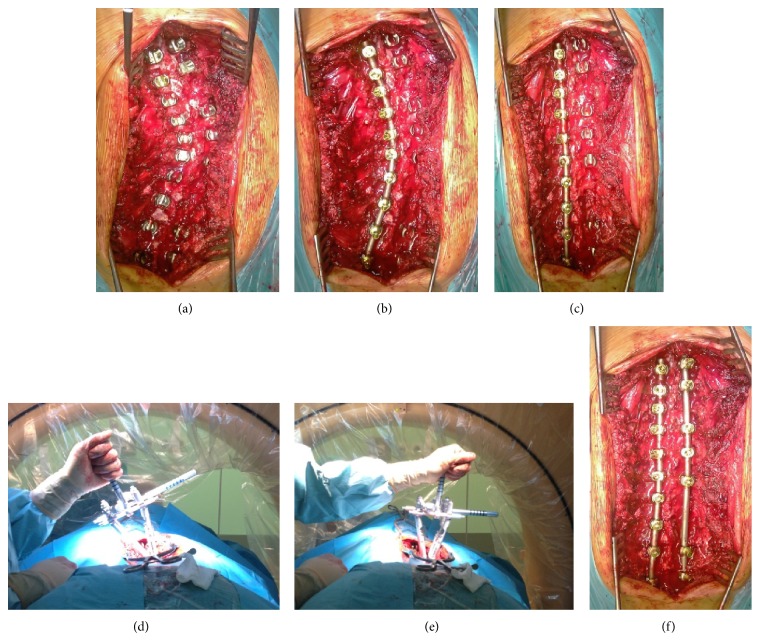
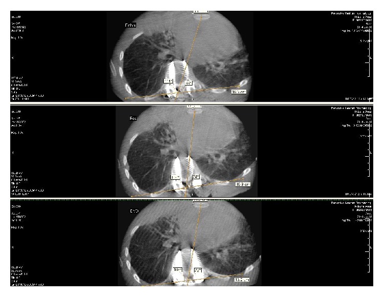
Numerous indirect methods for apical vertebral rotation (AVR) measurement have been reported and none of them seems to be as accurate as computed tomography evaluation. The aim of this study was to compare spinal rotation changes during innovative technique of intraoperative computed tomography (ICT) evaluation with indirect methods such as Perdriolle and clinical evaluation with scoliometer. We examined 42 adolescent idiopathic scoliosis (AIS) patients treated with posterior scoliosis surgery (PSS). The mean age at the time of surgery was 16 years. ICT evaluation was performed before and after scoliosis correction in prone position. Clinical rib hump measure with scoliometer and radiographic Perdriolle were performed before and after surgery. There was 71,5% of average rib hump correction with scoliometer but only 31% of correction with ICT (P=0,026) and there was no significant correlation between them (R=0,297, p=0,26). Mean postcorrectional Perdriolle AVR had a decrease of 16,5°. The average ICT AVR had a decrease of only 1,2° (P=0,003). There was no significant statistic correlation between ICT and Perdriolle AVR evaluation (R=0,297, p=0,2). There is a significant discrepancy in AVR and rib hump assessment between scoliometer and Perdriolle methods and ICT evaluation, which seems to be the most accurate tool for spinal derotation measurement.


Figures

Figure 1
Figure 1
Steps of the surgical correction, (a) screw placement, (b) rod contoured to the curve, (c) after “derotation on the rod,” (d) before DVD, (e) after DVD, and (f) second rod on the convex side.
Figure 2
Figure 2
(a) “Patient 2:” (A) pre- and (B) postoperative AFBT with Bunnell scoliometer. Standing position (C) before and (D) after correction. (b) “Patient 27:” (A) pre- and (B) postoperative AFBT. Standing position (C) before and (D) after correction.
Figure 3
Figure 3
(a) Perdriolle torsionmeter, (b) pre- and (c) postcorrectional Cobb angle measurement on plain radiographs.
Figure 4
Figure 4
Intraoperative computed tomography evaluation with O-Arm (Medtronic).
Figure 5
Figure 5
ICT evaluation AVR measure with Aaro and Dahlborn (A&D) method.
Figure 6
Figure 6
A new method of derotation assessment on the CT scans, two-step procedure. (a) Before correction, (b) after rod derotation, and (c) after DVD. Upper window: upper instrumented screw (UIS), middle window: apical screw (AS), and lower window: lower instrumented screw (LIS). The longitudinal axis of the particular screw was marked which crossed the middle of the head, the body, and tip of the screw. This line created an angle with the horizontal reference line. The difference of these angles between AS and UIS and AS and LIS before and after surgery allowed the real apical vertebral derotation (AVD) assessment. AS (°)-UIS (°): bigger windows in upper section, AS (°)-LIS (°): bigger windows in the lower section.
Figure 7
Figure 7
Rib hump angle ICT measurement (pleura-pleura (P-P) line and spine-sternum (S-S) line ratio). P-P line was marked between the two highest points of pleura at the apex of the curve. S-S line was marked between the middle of the basis of spinal process of apex vertebra with the midline of the sternum.
Figure 8
Figure 8
“Patient 13:” (a), (b), and (c) good clinical outcome. (d) Precorrectional CT scan of the same patient: upper window (UIS), middle (AS), lower window (LIS), AS  ( °) − UIS  ( °) = 35° (bigger window in upper section), AS  ( °) − LIS  ( °) = 13° (bigger window in the lower section). (e) Postcorrectional CT scan worsening of AVR after correction, AS  ( °) − UIS  ( °) = 34°, AS  ( °) − LIS  ( °) = 15°, AVR  precorrectional  ( °) = A&D  ( °) = 26°, AVD = (34° ± 15°)/2 − (35° ± 13°)/2 = 0,5°, AVR postcorrectional (°) = 26° + 0,5° = 26,5°. Poor ICT outcome in contrast with Perdriolle AVR.

References

    1. Cobb J. R. Outlines for the study of scoliosis. Instructional Course Lectures. 1948:261–275.
    1. Abul-Kasim K., Karlsson M. K., Hasserius R., Ohlin A. Measurement of vertebral rotation in adolescent idiopathic scoliosis with low-dose CT in prone position—method description and reliability analysis. Scoliosis Journal. 2010;5(1, article 4) doi: 10.1186/1748-7161-5-4. - DOI - PMC - PubMed
    1. Pankowski R., Roclawski M., Smoczynski A., et al. he influence of the development of scoliosis posterior surgery on 3-planar deformity correction. Research into Spinal Deformities. 2012;8:p. 492.
    1. Nash C. L., Jr., Moe J. H. A study of vertebral rotation. The Journal of Bone & Joint Surgery—American Volume. 1969;51(2):223–229. - PubMed
    1. Perdriolle R., Vidal J. Morphology of scoliosis: three-dimensional evolution. Orthopedics. 1987;10(6):909–915. - PubMed

Publication types

LinkOut - more resources