. 2015 Jun;21(6):1096-109.
doi: 10.1261/rna.048298.114. Epub 2015 Apr 10.

oskar RNA plays multiple noncoding roles to support oogenesis and maintain integrity of the germline/soma distinction

Affiliations

oskar RNA plays multiple noncoding roles to support oogenesis and maintain integrity of the germline/soma distinction

Matt Kanke et al. RNA. 2015 Jun.

Abstract

The Drosophila oskar (osk) mRNA is unusual in that it has both coding and noncoding functions. As an mRNA, osk encodes a protein required for embryonic patterning and germ cell formation. Independent of that function, the absence of osk mRNA disrupts formation of the karyosome and blocks progression through oogenesis. Here we show that loss of osk mRNA also affects the distribution of regulatory proteins, relaxing their association with large RNPs within the germline, and allowing them to accumulate in the somatic follicle cells. This and other noncoding functions of the osk mRNA are mediated by multiple sequence elements with distinct roles. One role, provided by numerous binding sites in two distinct regions of the osk 3' UTR, is to sequester the translational regulator Bruno (Bru), which itself controls translation of osk mRNA. This defines a novel regulatory circuit, with Bru restricting the activity of osk, and osk in turn restricting the activity of Bru. Other functional elements, which do not bind Bru and are positioned close to the 3' end of the RNA, act in the oocyte and are essential. Despite the different roles played by the different types of elements contributing to RNA function, mutation of any leads to accumulation of the germline regulatory factors in the follicle cells.

Keywords: Bruno; lncRNA function; oskar; protein sequestration.


Figures

FIGURE 1.
FIGURE 1.
Bru binding sites are required for osk RNA function. (A) Schematic of the osk 3′ UTR indicating the locations of Bru binding sites (BREs, type II and type III). The sites are clustered in the AB and C regions. (B) Rescue of the osk RNA null egg laying defect by osk transgenes. Rates of egg laying (see Materials and Methods) for females lacking endogenous osk mRNA [oskA87/Df(3R)osk] but carrying a single copy of an osk transgene, as indicated. The rate obtained with a single copy of the osk+ transgene was set at 100%. (C) Transcript levels for osk transgenes. The rp49 mRNA was used as a control to ensure that similar amounts of ovarian RNA were used for each genotype. (D) Addition of Bru binding sites rescues the egg laying defect of osk ABC BRE. In all cases females lack endogenous osk mRNA and carry the nosGAL4::VP16 driver and a single copy of the osk ABC BRE transgene, which provides only partial rescue of egg laying. A UAS-GFP transgene or UAS-GFP-4xBRE transgene was also present, as indicated. (E) Reducing Bru activity partially suppresses the oogenesis progression defect of osk RNA null females. Median lengths of developmentally arrested egg chambers were measured for the genotypes shown (n for oskA87/Df(3R)pXT103, 16; with aretPA/+, 37; with aretQB/+, 36). P values derived from the Kolmogorov–Smirnov Test: (*) P < 0.05; (**) P < <0.01. (F) Reducing Bru activity rescues the egg laying defect of osk ABC BRE−. Rates of egg laying for females lacking endogenous osk mRNA [oskA87/Df(3R)osk] but carrying an osk+ or osk ABC BRE transgene were determined, testing the consequences of reducing Bru activity by heterozygosity for aretz2286. (GI) Ovarioles from wild type (G), oskA87/Df(3R)osk (H), or oskA87/Df(3R)osk expressing the osk C all transgene (I). The ovarioles were stained with ToPro for DNA (red) and anti-Hts for Adducin-like (green). (J) UV crosslinking assay of Bru in ovarian extract binding to osk RNA probes. The AB and C regions contain the Bru binding sites, and are as previously defined (Kim-Ha et al. 1995). Deletion of part of the C region enhances Bru binding, perhaps by altering secondary structure that would otherwise limit accessibility. Even with the enhanced binding, the C region binds substantially less Bru than does the AB region. Similar amounts of each probe were used. All lanes are from the same autoradiogram of a single experiment and gel, with irrelevant lanes removed.
FIGURE 2.
FIGURE 2.
Mapping regions of the osk 3′ UTR that contribute to osk RNA function. (A) The osk 3′ UTR is shown in schematic form, with regions included in transgenes shown as horizontal gray bars. Defined Bru binding sites are indicated by black rectangles and the OES (the signal that mediates transport of the mRNA to the oocyte) is indicated by a red bar. The K10-TLS, the oocyte entry signal of fs(1)K10, is indicated by a blue bar. The osk sequences were incorporated into UAS-egfp transgenes, except for K10-TLS 119 of 3b, which lacks gfp (Materials and Methods). Each transgene was tested in the oskA87/Df(3R)pXT103 background with the pCog-Gal4:VP16 (Rørth 1998) and nanos-Gal4:VP16 (Van Doren et al. 1998) drivers. Results of the assays are indicated at right. RNA enriched: +, strong enrichment of the RNA in the oocyte; −, no enrichment in the oocyte. For the complementation test the distributions of the two mRNAs were not monitored directly (n.a.), but can be inferred from tests with the individual mRNAs. RNA null rescue: +, eggs laid; −, no eggs laid. The presence of eggs was scored, not the frequency of egg laying. (B) Representative in situ hybridizations against the GFP portion of the transgenic construct. Transgenic RNA signal in red and DNA in blue (scale bar, 30 µm).
FIGURE 3.
FIGURE 3.
Fine scale mapping of osk RNA function elements. (A) Mutations in the osk 3′ region. The sequence of the region is shown, with Bru binding sites (BREs, type II, and type III sites) marked in blue. Black bars above the sequence indicate minimal fragments tested in Figure 2 (the final 18 nt of the osk 3′ UTR are not shown, but are present in the minimal fragments along with a further 8 nt of genomic DNA). Beneath the sequence are shown the mutations (lowercase) introduced into genomic osk transgenes. The osk CII mutant has the mutations of both osk 3920-923 and osk 3970-974. Single copies of each transgene were tested in the oskA87/Df(3R)osk background for rate of egg laying and mRNA level. At least two independent transgenic lines were tested for mutants with substantial defects. The additional lines of mutants osk3977-981 and osk3984-988 also lacked detectable activity. RNA levels were determined as in Figure 1, using rp49 as an internal control. (B) Affinity capture assay of Bru binding to osk C region RNAs, either wild type or with scanning mutations. The osk RNAs are fused to the S1 aptamer (the first two lanes are the aptamer alone). After incubating with ovary extract, the RNAs and bound proteins were recovered by affinity purification to generate supernatent (S; unbound) and pellet (P; bound) fractions, and the presence of Bru was determined by Western blotting. How well each version of osk mRNA supports the osk RNA function is indicated at bottom. (C) Increasing transgene dosage to raise osk mRNA levels for selected mutants. The RNase protection assays are shown at left with transgenic osk mRNAs indicated (all were in the oskA87/Df(3R)osk background), and the quantitation by phosphorimaging at right (samples in the same order). (D,E) Ovarioles stained with TOPRO-3 to detect nuclei. Both are oskA87/Df(3R)osk, with E expressing two copies of the osk3977-981 transgene (same genotype as in panel C). (F) Distribution of mutant osk mRNAs. All egg chambers are from oskA87/Df(3R)osk females expressing a single copy of the transgene indicated. osk mRNA (green) was detected by in situ hybridization, with DNA (red) labeled with DAPI.
FIGURE 4.
FIGURE 4.
Karyosome defects of osk RNA null mutants. (A) Suppression of karysome defects by reducing aret activity. Females are all oskA87/Df(3R)pXT103, with the aret al.leles indicated at top. (B) Frequency of karyosome defects for osk mutants. Egg chambers from oskA87/Df(3R)osk females expressing a single copy of the transgene indicated were scored for karyosome morphology (for osk ABC BRE, the females were osk0/Df(3R)osk). Mutants osk3889-893, osk3896-900, osk3903-907, osk3909-913, osk3915-919, osk3920-923, osk3950-954, osk3957-961, and osk3963-967, which are not included in the diagram, all had 100% normal karyosomes with n values of 9 or greater. Two independent transgenic lines were tested for the scanning mutants shown. To facilitate comparison, the results of the egg laying tests (Figs. 1B, 3A) are summarized below the graph. (C) Examples of karyosome morphology. Complete egg chambers are shown for examples of wild type (w1118) and oskA87/Df(3R)osk (left and center images in the top row). For the other panels only the oocyte is shown. Samples were labeled with TOPRO-3 for DNA (red) and anti-lamin (green).
FIGURE 5.
FIGURE 5.
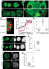
Redistribution of Bru and other germline proteins in osk RNA null ovaries. (AD) Immunodetection of Bru in wild-type (A,B) and osk RNA null (osk0/osk0) ovaries (C,D). The higher magnification samples (B,D) show the punctate distribution of Bru, which is reduced in the osk RNA null mutant (see also Fig. 6). The level of Bru is much greater in the oocyte than the nurse cells, as can be seen in egg chambers in which the confocal section includes the oocyte (examples in A,C,D). (E) Organization of the egg chamber. Half of an egg chamber is shown, with the different cell types indicated at left. To monitor Bru signal in nurse cells and follicle cells, signal intensity was measured along multiple lines (e.g., the white lines) in Fiji (Materials and Methods). (F) Traces of signal intensity along four lines are shown for both wild-type and osk RNA null egg chambers. The relative positions of follicle and nurse cells along the lines are indicated below. (G) Quantitation of Bru signal intensity in follicle cells. Each data point represents a region crossing multiple follicle cells (thus including a mixture of nuclei and cytoplasm) and avoiding the inner (adjacent to the germline cells) and outer boundaries of the follicle cell layer. The “rescue” sample was osk0/osk0 with an osk transgene providing full rescue of the oogenesis arrest phenotype. (H,I) Immunodetection of Orb in wild-type and osk RNA null ovaries. For H′ and I′, signal intensities were adjusted identically in Photoshop to enhance the signal. (J) Quantitation of Orb signal intensity in areas of follicle cells, as in G. The difference is statistically significant: from an unpaired two-tailed Student's t test, P < 0.001. (K,L) Immunodetection of CG9925 in wild-type and osk RNA null ovaries. For K′ and L′, signal intensities were adjusted identically in Photoshop to enhance the signal and make the follicle cell patterns more visible. Note that the highly punctate distribution of CG9925 protein precludes the type of analysis shown in panel F. (M) Quantitation of CG9925 signal intensity in areas of follicle cells, as in G. The difference is statistically significant: from an unpaired two-tailed Student's t test, P < 0.001.
FIGURE 6.
FIGURE 6.
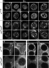
Loss of Bru and Orb from large cytoplasmic RNPs in osk RNA null ovaries. (A) Immunodetection of Bru in early-stage egg chambers, either wild type (w1118; upper two rows) or osk RNA null (osk0/osk0; lower two rows). In wild type, Bru is strongly enriched in perinuclear nuage. In the upper left panel of each set a nurse cell nucleus is indicated with an arrowhead to show enriched Bru in nuage, or an arrow to show the substantially reduced level of Bru in nuage in the absence of osk mRNA. This pattern is highly consistent, as seen in the 10 panels for each genotype. Note that some osk RNA null egg chambers have excess nurse cells. Each egg chamber in panels marked with a white asterisk has >10 germline cell nuclei in the single focal plane of the image. The rightmost of these examples also has two germline cells highly enriched with Bru, indicating that two cells are developing as oocytes. (B) Higher magnification views to show both perinuclear nuage and sponge bodies. As in A, nuclei are marked with arrowheads to show perinuclear nuage with enriched Bru, or arrows to show the loss of Bru from nuage. Arrowheads also indicate examples of Bru in cytoplasmic puncta, corresponding to sponge bodies (Snee and Macdonald 2009). (C) Enrichment of Orb in nuage in wild type (arrowheads), but not in osk RNA null egg chambers (arrows).
FIGURE 7.
FIGURE 7.
Mutants defective in osk RNA function fail to rescue the Bru redistribution phenotype. (AF) Immunodetection of Bru. Genotypes are shown at top for A and B. For panels CF, the osk transgenes (indicated at top) are in the oskA87/Df(3R)osk background. (G) Quantitation of signal intensity in areas of follicle cells, as in Figure 5.

Similar articles

Cited by

References

    1. Bartel DP 2009. MicroRNAs: target recognition and regulatory functions. Cell 136: 215–233. - PMC - PubMed
    1. Bohrmann J, Haas-Assenbaum A 1993. Gap junctions in ovarian follicles of Drosophila melanogaster: inhibition and promotion of dye-coupling between oocyte and follicle cells. Cell Tissue Res 273: 163–173. - PubMed
    1. Cech TR, Steitz JA 2014. The noncoding RNA revolution—trashing old rules to forge new ones. Cell 157: 77–94. - PubMed
    1. Ephrussi A, Dickinson LK, Lehmann R 1991. oskar organizes the germ plasm and directs localization of the posterior determinant nanos. Cell 66: 37–50. - PubMed
    1. Furriols M, Casanova J 2014. Germline and somatic vitelline proteins colocalize in aggregates in the follicular epithelium of Drosophila ovaries. Fly (Austin) 8: 113–119. - PMC - PubMed

Publication types

LinkOut - more resources