. 2015 Apr 10;4(5):636-48.
doi: 10.1242/bio.201511551.

Loss of a Clueless-dGRASP complex results in ER stress and blocks Integrin exit from the perinuclear endoplasmic reticulum in Drosophila larval muscle

Affiliations

Loss of a Clueless-dGRASP complex results in ER stress and blocks Integrin exit from the perinuclear endoplasmic reticulum in Drosophila larval muscle

Zong-Heng Wang et al. Biol Open. .

Abstract

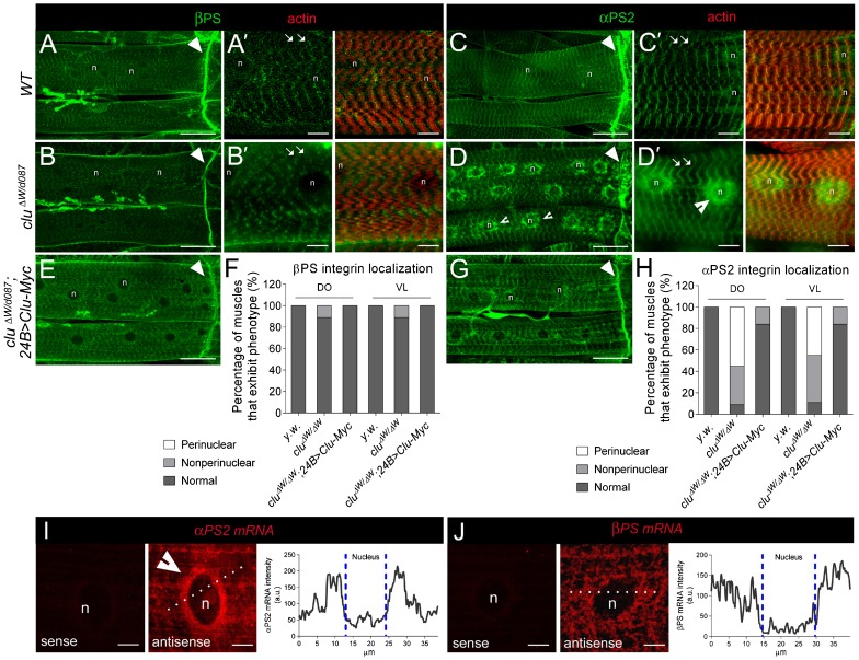
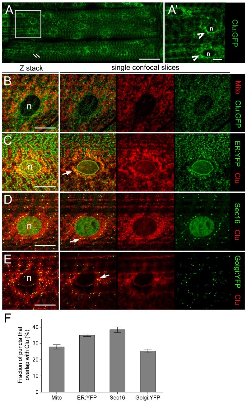
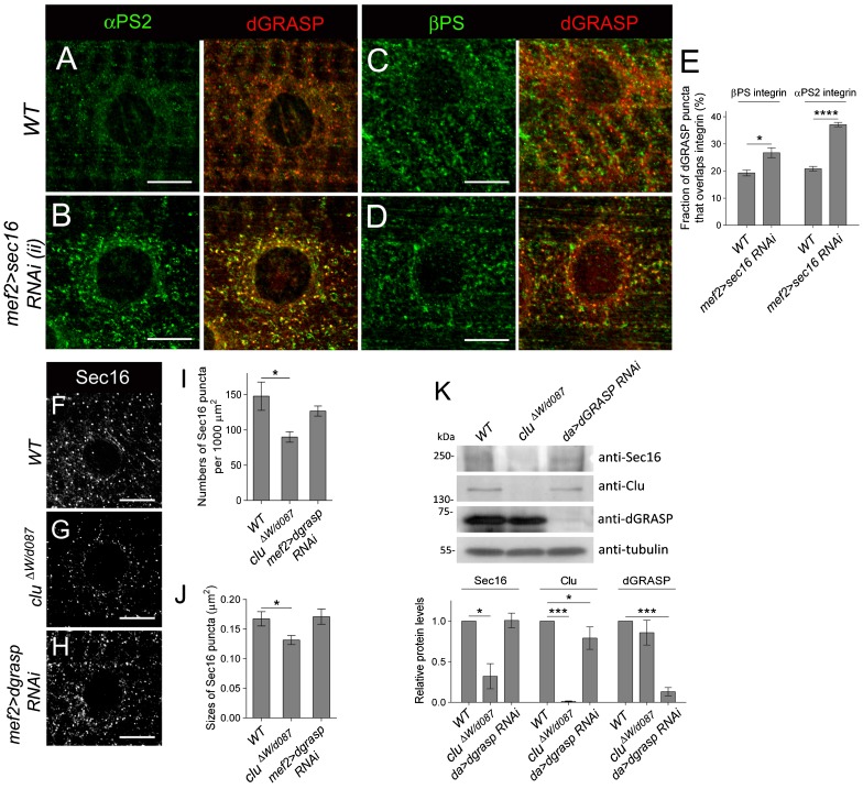
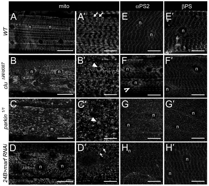
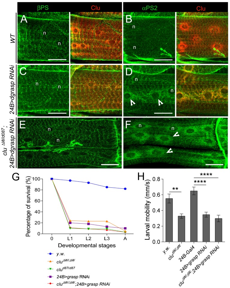
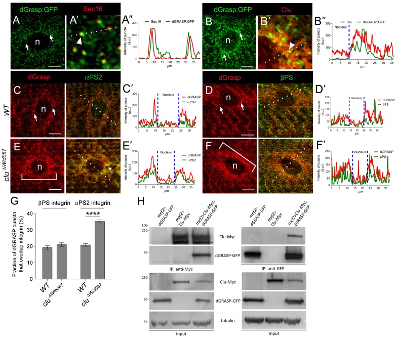
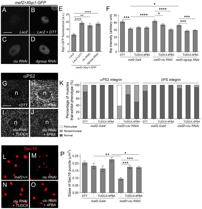
Drosophila Clueless (Clu) and its conserved orthologs are known for their role in the prevention of mitochondrial clustering. Here, we uncover a new role for Clu in the delivery of integrin subunits in muscle tissue. In clu mutants, αPS2 integrin, but not βPS integrin, abnormally accumulates in a perinuclear endoplasmic reticulum (ER) subdomain, a site that mirrors the endogenous localization of Clu. Loss of components essential for mitochondrial distribution do not phenocopy the clu mutant αPS2 phenotype. Conversely, RNAi knockdown of the Drosophila Golgi reassembly and stacking protein GRASP55/65 (dGRASP) recapitulates clu defects, including the abnormal accumulation of αPS2 and larval locomotor activity. Both Clu and dGRASP proteins physically interact and loss of Clu displaces dGRASP from ER exit sites, suggesting that Clu cooperates with dGRASP for the exit of αPS2 from a perinuclear subdomain in the ER. We also found that Clu and dGRASP loss of function leads to ER stress and that the stability of the ER exit site protein Sec16 is severely compromised in the clu mutants, thus explaining the ER accumulation of αPS2. Remarkably, exposure of clu RNAi larvae to chemical chaperones restores both αPS2 delivery and functional ER exit sites. We propose that Clu together with dGRASP prevents ER stress and therefore maintains Sec16 stability essential for the functional organization of perinuclear early secretory pathway. This, in turn, is essential for integrin subunit αPS2 ER exit in Drosophila larval myofibers.

Keywords: Clueless; Drosophila; Integrin; Muscle; Trafficking; dGRASP.


Conflict of interest statement

Competing interests: The authors declare no competing or financial interests.

Figures

Fig. 1.
Fig. 1.. αPS2 accumulates within contractile muscles upon loss of Clu.
(A–E,G) Immunolocalization of integrin proteins (green) and F-actin (red; phalloidin) in muscles of filleted L3 individuals (n = nucleus). (A–B′) In both WT and clu/ muscles, βPS integrin is found at MASs (arrowheads in A,B) and costamere structures that encircle the sarcolemma along the length of the muscle (arrows in A′,B′). (C,C′) αPS2 integrin also accumulates at the ends of WT muscles (arrowhead in C) and at costameres (arrows in C′). (D,D′) In clu mutants, the αPS2 subunit accumulates around the periphery of the nucleus, as indicated by the indented arrowheads. (E,G) The reintroduction of full-length clu cDNA into clu mutant muscle tissue has no effect on βPS integrin distribution (E) and restores the accumulation of αPS2 to its normal location within the cell (G). (F,H) Quantification of βPS and αPS2 integrin distribution in the dorsal oblique (DO; 16<n<36) and ventral longitudinal (VL; 16<n<36) L3 muscles of indicated the genotypes. (I,J) Fluorescent in situ hybridizations (FISH) in L3 muscle tissue. αPS2 mRNA accumulates around the nuclei (I, middle panel; n = 42), while βPS mRNA appears evenly distributed throughout the muscle cell (J; n = 23). The sense probes for both mRNAs reveal little background signal (left panels). Quantitation of fluorescence intensity (dotted line) shows that the perinuclear signal of αPS2 mRNA is higher than that of βPS2 mRNA. Scale bars, 50 µm (A–E,G), 10 µm (A′–D′), 5 µm (I,J).
Fig. 2.
Fig. 2.. Clu localizes with dGRASP and Sec16 ER exit sites.
(A–G) Muscle tissue from L3 larvae was dissected and immunostained to examine the subcellular localization of Clu protein. (A,A′) The Clu:GFP protein trap line (green) localizes in a repeated pattern within the muscle (arrows) and accumulates around the nuclei (n; indented arrows). The right panel is a close up of the boxed region in the left panel. (B) The perinuclear accumulation of Clu:GFP (green) reveals little colocalization with mitochondria (red; anti-Complex V). (C–F) An anti-Clu antibody (red) was used to confirm the Clu:GFP nuclear staining pattern and also to discern the localization of Clu puncta with other organelle markers (green) in WT larval muscle. A composite Z-stack is followed by representative single confocal slices. (C,D) Clu-positive puncta overlap with both a general ER marker (C) and the ERES protein Sec16 (D). (E,F) Clu colocalizes with a subset of Golgi:YFP puncta (E). (F) The percentage of puncta of each organelle marker that overlap with Clu protein. Colocalization was determined from multiple single plane images calculated using the Image J JACoP plug-in. Mean±s.e.m. Scale bars, 50 µm (A), 10 µm (A′,B–G).
Fig. 6.
Fig. 6.. Sec16 protein levels are reduced in clu mutants.
(A–D) Immunostaining of integrin (green) and dGRASP (red) in L3 contractile muscles. Low amounts of both αPS2 (A) and βPS (C) colocalize with dGRASP around nuclei (n) in WT muscle cells. (B,D) RNAi knockdown of Sec16 in muscle tissues results in the retention of αPS2 in dGRASP-positive puncta (B), while low levels of βPS accumulate around the nuclei (D). (E) Graph depicting the fraction of Sec16 puncta that overlap integrins based upon analysis of multiple images like those presented in panels A–F (*p<0.05; ***p<0.0005). (F–H) Perinuclear staining of Sec16 staining in the indicated genotypes. Sec16 puncta are reduced in clu mutants (G) when compared to WT (F) or dgrasp-depleted muscle tissue (H). (I,J) The number (I) and size (J) of Sec12-positive ERES are reduced in clu mutants. (K) Western blot and band intensity quantification of Sec16, Clu and dGRASP protein levels in the indicated genotypes. Sec16 protein levels are reduced in clu, but not dGRASP mutants (mean±s.e.m.; *p<0.05; ***p<0.005). Scale bars, 10 µm (A–D,F–H).
Fig. 3.
Fig. 3.. The requirement for Clu and dGRASP in integrin localization is separable from mitochondrial organization.
(A–D) The distribution of mitochondria (anti-Complex V) in muscle 6 (n = nucleus). (A–D) Z-stacks of the muscle surface and internal myofibrils. (A′–D′) Internal muscle cell slices. (A,A′) The mitochondria in WT muscle cells are evenly distributed and align in a repeated pattern (arrows). (B–D′) Either clu/ (B,B′) or parkin/− (C,C′) mutants exhibit severe mitochondrial clustering (arrowheads). Knockdown of marf RNAi in the muscle with 24B-GAL4 results in fragmented mitochondria (small arrows). (E–H) The perinuclear αPS2 localization phenotype is only apparent in clu mutants (indented arrows in F), and not upon a decrease in parkin (G) or marf (H). (E′–H′) βPS does not accumulate in the perinuclear region in WT (E′), clu/ (F′) mutants, parkin/ mutants (G′), or marf RNAi muscles (H′). Scale bars, 50 µm (A–D,G), 10 µm (E–H, A′–H′).
Fig. 4.
Fig. 4.. dGRASP RNAi in the muscle phenocopies clu mutants.
(A–D) L3 muscle fillets reveal the localization of integrins (green) and Clu (red). βPS (A) and αPS2 (B) show relatively normal integrin distribution in WT muscle (n = nucleus). In 24B>dgrasp RNAi muscles, βPS integrin appears WT (C), while the αPS2 subunit colocalizes with endogenous Clu in the nuclear periphery (D; indented arrowheads). (E,F) βPS localization in muscles mutant for clu that also knockdown dGRASP levels (cluΔW/clud087; 24B> dgrasp RNAi) are similar to WT (E), while the perinuclear distribution of αPS2 looks like clu/ or dgrasp RNAi alone (F; indented arrowheads). (G) Survival curve for clu and dgrasp mutants at different developmental stages (E, embryo; L1, 1st instar larva; L2, 2nd instar larva; 3rd instar larva; A, adult). (H) Locomotor activity analysis for early L3 larvae of indicated genotypes (mean±s.e.m.; **p<0.005; ****p<0.0001). Scale bars, 25 µm (A–D); 50 µm (E,F).
Fig. 5.
Fig. 5.. Clu physically binds to and mediates the localization of dGRASP to puncta.
(A–B″) UAS-dGRASP-GFP is expressed in the muscle using by mef2-GAL4 and is found in puncta surrounding the nuclei (n; arrows in A,B). High magnification images and line intensity profiles (dotted lines) reveal a partial overlap with Sec16 (arrowheads in A′,A″) and colocalization with Clu (arrowheads in B′,B″). (C–F′) Micrographs (C–F) and the corresponding fluorescent intensity line profiles to illustrate colocalization (C′–F′; dotted lines) between dGRASP (red) and integrins (green). The dGRASP-positive puncta (arrows) at the ERES and Golgi exhibit little overlap with αPS2 (C,C′) and βPS (D,D′) in the cell. dGRASP protein is more diffuse in clu mutants (E,F; brackets) and colocalizes with αPS2 around the nuclei (asterisk in E,E′). (G) Quantitation of results in panels C–F showing the fraction of dGRASP signal that colocalizes with integrins (mean±s.e.m.; ****p<0.0001). (H) A myc-tagged version of Clu and a dGRASP-GFP fusion protein were expressed using the GAL4/UAS system in the L3 stage. Immunoprecipitation of the resulting lysates with either anti-myc (left panel) or anti-GFP (right panel) resulted in the detection of a Clu-dGRASP complex using Western blot analysis. Asterisk indicates background band. Scale bars, 5 µm (A–F).
Fig. 7.
Fig. 7.. Molecular chaperones can alleviate ER stress due to a reduction in either Clu or dGRASP.
(A–F) ER stress markers are upregulated in clu or dGRASP RNAi. The ER stress reporter Xbp1-GFP is elevated upon induction of ER stress by DTT (B) or upon RNAi knockdown of clu (C) or dGRASP (D) in L3 muscles (n = nucleus). (E) Quantitation of the ER stress inducer Xbp1-GFP in the indicated genotypes. (F) Independent measurements of ER stress measuring the amount of Bip levels in L3 muscle. ER stress in increased upon feeding with DTT or in clu or dGRASP RNAi and can be ameliorated upon treatment with the molecular chaperones TUDCA or 4PBA (*p<0.05; ***p<0.0005; ****p<0.0001). (G–K) αPS2 accumulates in clu RNAi (H) muscle tissue and this perinuclear accumulation is alleviated upon treatment with TUDCA (I) or 4PBA (J). (K) Graph depicting the internal accumulation of αPS2 upon loss of Clu only. (L–P) The size of Sec16 ERES is reduced in clu RNAi (M), but is restored upon inhibition of ER stress (N–P) (mean±s.e.m.; *p<0.05; **p<0.01; ***p<0.005). Scale bars, 10 µm (A–D, G–J); 2 µm (L–O).

Similar articles

Cited by

References

    1. Baehrecke E. H. (1997). who encodes a KH RNA binding protein that functions in muscle development. Development 124, 1323–1332. - PubMed
    1. Barlowe C. K., Miller E. A. (2013). Secretory protein biogenesis and traffic in the early secretory pathway. Genetics 193, 38 10 10.1534/genetics.112.142810 - DOI - PMC - PubMed
    1. Barr F., Puype M., Vandekerckhove J., Warren G. (1997). GRASP65, a protein involved in the stacking of Golgi cisternae. Cell 91, 253–262. - PubMed
    1. Behnia R., Barr F. A., Flanagan J. J., Barlowe C., Munro S. (2007). The yeast orthologue of GRASP65 forms a complex with a coiled-coil protein that contributes to ER to Golgi traffic. J. Cell Biol. 176, 255–261. 10.1083/jcb.200607151 - DOI - PMC - PubMed
    1. Bhattacharyya D., Glick B. S. (2007). Two mammalian Sec16 homologues have nonredundant functions in endoplasmic reticulum (ER) export and transitional ER organization. Mol. Biol. Cell 18, 839–849. 10.1091/mbc.E06-08-0707 - DOI - PMC - PubMed