Review
. 2015 Dec;124(4):443-62.
doi: 10.1007/s00412-015-0514-0. Epub 2015 Apr 11.

New insights into the mechanism of DNA mismatch repair

Affiliations
Review

New insights into the mechanism of DNA mismatch repair

Gloria X Reyes et al. Chromosoma. 2015 Dec.

Abstract

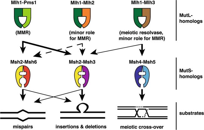
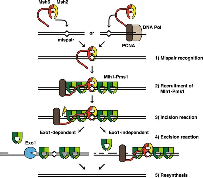
The genome of all organisms is constantly being challenged by endogenous and exogenous sources of DNA damage. Errors like base:base mismatches or small insertions and deletions, primarily introduced by DNA polymerases during DNA replication are repaired by an evolutionary conserved DNA mismatch repair (MMR) system. The MMR system, together with the DNA replication machinery, promote repair by an excision and resynthesis mechanism during or after DNA replication, increasing replication fidelity by up-to-three orders of magnitude. Consequently, inactivation of MMR genes results in elevated mutation rates that can lead to increased cancer susceptibility in humans. In this review, we summarize our current understanding of MMR with a focus on the different MMR protein complexes, their function and structure. We also discuss how recent findings have provided new insights in the spatio-temporal regulation and mechanism of MMR.


Figures

Fig. 1
Fig. 1
S. cerevisiae MSH and MLH complexes. The arrows indicate their functional interactions and potential functions in vivo. Arrows denote major roles (thick arrows) and minor roles (thin arrows) in the indicated processes. Dashed lanes represent interactions that are biologically relevant apparently only in specific genetic backgrounds
Fig. 2
Fig. 2
Crystal structure of E. coli MutS and human MutSα according to previous reports (Lamers et al. 2000; Warren et al. 2007). a MutS homodimer, in which the mispair-contacting subunit has been colored by domain (I–VI) with red, yellow, green, purple, pink, and brown, respectively. DNA (blue sticks) and ADP are also indicated. b Human Msh2-Msh6 complex. Msh2 is shown in yellow and Msh6 in red. c Expanded view of the region in the black box in (b) that directly interacts with the mispaired base. The conserved Msh6 residues phenylalanine (F432) and glutamic acid 434 (E434), as well as the mispaired bases guanine (G) and thymine (T) are indicated
Fig. 3
Fig. 3
Structural domains of MutL and eukaryotic MutL homologs. The N-terminal domain (NTD) is indicated by a gray box that contains an ATPase domain consisting of four highly conserved motifs (shown in red). The dimerization domain and the endonuclease motif DQHA(X)2E(X)4E (light blue box) contained within the C-terminal domain (CTD) (blue box) are indicated. The conserved FERC sequence of Mlh1 is shown in yellow
Fig. 4
Fig. 4
Model for the Mlh1-Pms1 complex in S. cerevisiae adapted from Smith et al. 2013. a Mlh1 is shown in dark green and Pms1 in brighter green. Illustration of the N-terminal domains was generated based on structure pdb.3H4L (Arana et al. 2010), whereas the C-terminal domain structure corresponds to pdb.4e4w (Gueneau et al. 2013). N- and C-terminal domains are linked by an unstructured linker (dashed line). b Expanded view of the conserved amino acids comprising the metal binding pocket (metal ions in black) of the composite endonuclease site located within the black box in (a)
Fig. 5
Fig. 5
Model of alternative excision pathways that act during MMR adapted from Goellner et al. 2014. The diagram describes the sequential steps of the MMR reaction. The Msh2-Msh6 heterodimer recognizes mispairs (1). A fraction of the recognition complexes is coupled to replication machinery (PCNA-DNA Pol) through PCNA. Alternatively, the Msh2-Msh6 complex scans the DNA for mispairs in a PCNA-interaction independent manner. The mispair-bound Msh2-Msh6 complex recruits Mlh1-Pms1 (2). The loading of Mlh1-Pms1 onto DNA is catalytic whereby one mispair recognition complex recruits several Mlh1-Pms1 complexes to or near the mispair site. The Mlh1-Pms1 endonuclease is then activated by PCNA to nick the DNA comprising the incision step (3). At this step, Msh6 plays a role by retaining or recruiting PCNA in proximity to the mispair site. The excision reaction (4) can be mediated by Exo1 (Exo1-dependent) or by unknown factors (Exo1-independent) potentially including multiple rounds of incision by Mlh1-Pms1, strand displacement by the DNA replication machinery or other exonucleases followed by resynthesis of the DNA (5)

References

    1. Acharya S, Wilson T, Gradia S, Kane MF, Guerrette S, Marsischky GT, Kolodner R, Fishel R. hMSH2 forms specific mispair-binding complexes with hMSH3 and hMSH6. Proc Natl Acad Sci U S A. 1996;93:13629–13634. - PMC - PubMed
    1. Acharya S, Foster PL, Brooks P, Fishel R. The coordinated functions of the E. coli MutS and MutL proteins in mismatch repair. Mol Cell. 2003;12:233–246. - PubMed
    1. Ahrends R, Kosinski J, Kirsch D, Manelyte L, Giron-Monzon L, Hummerich L, Schulz O, Spengler B, Friedhoff P. Identifying an interaction site between MutH and the C-terminal domain of MutL by crosslinking, affinity purification, chemical coding and mass spectrometry. Nucleic Acids Res. 2006;34:3169–3180. - PMC - PubMed
    1. Allen DJ, Makhov A, Grilley M, Taylor J, Thresher R, Modrich P, Griffith JD. MutS mediates heteroduplex loop formation by a translocation mechanism. EMBO J. 1997;16:4467–4476. - PMC - PubMed
    1. Allen-Soltero S, Martinez SL, Putnam CD, Kolodner RD. A Saccharomyces cerevisiae RNase H2 interaction network functions to suppress genome instability. Mol Cell Biol. 2014;34:1521–1534. - PMC - PubMed

Publication types