Review
. 2015 Sep;67(1):85-100.
doi: 10.1016/j.molimm.2015.03.245. Epub 2015 Apr 8.

MBL-associated serine proteases (MASPs) and infectious diseases

Affiliations
Review

MBL-associated serine proteases (MASPs) and infectious diseases

Marcia H Beltrame et al. Mol Immunol. 2015 Sep.

Abstract

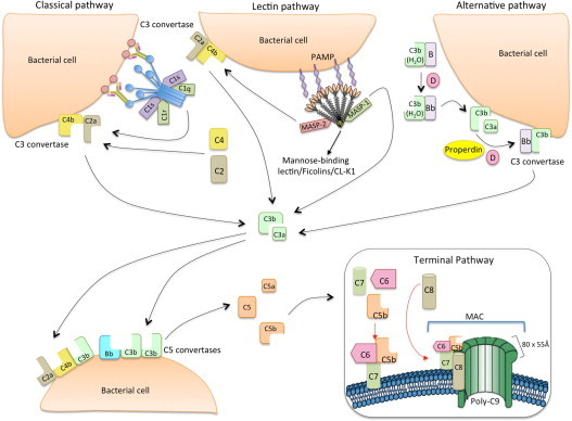
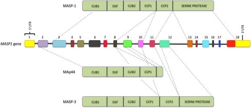
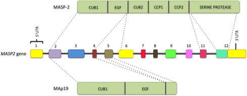
The lectin pathway of the complement system has a pivotal role in the defense against infectious organisms. After binding of mannan-binding lectin (MBL), ficolins or collectin 11 to carbohydrates or acetylated residues on pathogen surfaces, dimers of MBL-associated serine proteases 1 and 2 (MASP-1 and MASP-2) activate a proteolytic cascade, which culminates in the formation of the membrane attack complex and pathogen lysis. Alternative splicing of the pre-mRNA encoding MASP-1 results in two other products, MASP-3 and MAp44, which regulate activation of the cascade. A similar mechanism allows the gene encoding MASP-2 to produce the truncated MAp19 protein. Polymorphisms in MASP1 and MASP2 genes are associated with protein serum levels and functional activity. Since the first report of a MASP deficiency in 2003, deficiencies in lectin pathway proteins have been associated with recurrent infections and several polymorphisms were associated with the susceptibility or protection to infectious diseases. In this review, we summarize the findings on the role of MASP polymorphisms and serum levels in bacterial, viral and protozoan infectious diseases.

Keywords: Complement system; MASP-1; MASP-2; MASP-3; MAp19; MAp44.


Figures

Fig. 1
Fig. 1
The classical, lectin and alternative pathways of complement activation. The classical pathway is initiated via binding of C1 complex (which consists of C1q, C1r and C1s molecules) through its recognition molecule C1q to target molecules on the surface of pathogens. Subsequently, C1s cleaves C4, which binds covalently to the pathogen surface, and then cleaves C2, leading to the formation of C4b2a complex, the C3 convertase of the classical pathway. Activation of the lectin pathway occurs through binding of mannose-binding lectin (MBL) oligomers, ficolin oligomers or collectin heteromers (CL-K1 + CL-L1), complexed with MBL-associated serine proteases 1 and 2 homodimers (MASP-1 and MASP-2, respectively), to various carbohydrates or acetylated groups on the surface of pathogens (PAMPs: pathogen associated molecular patterns). Like C1s, MASP-2 leads to the formation of the C3 convertase, C4b2a, but its activation is dependent on MASP-1. MASP-1 also cleaves C2. Activation of the alternative pathway depends on spontaneous low-grade hydrolysis of C3 in plasma leading to the formation of C3b. This C3b binds Factor B (homologous to C2) to form a C3bB complex. The cleavage of Factor B by Factor D form the alternative pathway C3 convertase, C3bBb. Properdin stabilizes this complex. The C3 convertases cleave C3 to C3b, which bind covalently next to the site of complement activation (opsonization). This amplifies the cascade and mediates phagocytosis, as well as adaptive immune responses. The addition of additional C3b molecules to the C3 convertase forms C5 convertases (C3bBbC3b for the alternative pathway or C4bC2aC3b for both classical and lectin pathways). This C3b acts as a binding site for C5 and initiate the assembly of the membrane-attack complex (MAC) by cleavage of C5–C5a and C5b. Whereas C5a acts as a potent anaphylatoxin, C5b forms a complex with C6 and C7, which is inserted in the cell membrane. Thereafter, C8 and 10 – 18 C9 molecules (80 × 55 Å each) bind to this complex, resulting in a fully functional MAC (C5b-9). The three pathways converge to this common terminal pathway, culminating with cell lysis and death (Abbas et al., 2012).
Fig. 2
Fig. 2
MASP1 gene and MASP-1, MAp44, and MASP-3 proteins. Exons are numbered and not depicted to scale. MASP: mannose-binding lectin associated serine protease. MAp44: mannose-binding lectin associated protein of 44 kDa. CUB: C1r/C1s, Uegf, and bone morphogenetic protein; EGF: epidermal growth factor; CCP: complement control protein.
Fig. 3
Fig. 3
MASP2 gene and the mannose-binding lectin associated serine protease 2 (MASP-2) and MAp19 proteins. Exons are numbered and not depicted in scale. CUB: C1r/C1s, Uegf, and bone morphogenetic protein; EGF: epidermal growth factor; CCP: complement control protein.
Fig. 4
Fig. 4
Dual role of complement proteins during Trypanosoma cruzi infection (Sánchez Valdéz et al., 2013). Epimastigotes, which are the flagellated forms in Triatominae vectors, are susceptible to complement-mediated lysis. Under natural conditions however, they are never confronted with it. On the other hand, the amastigote and trypomastigote forms infecting humans, resist complement-mediated lysis. Moreover, they use recognition by MBL/ficolins/MASP complexes to invade host cells. Several complement inhibitor molecules are expressed on T. cruzi surface, such as: Gp 58/68 (Fischer et al., 1988), Trypanosoma cruzi-decay accelerating factor (Norris et al., 1991), Complement regulator protein (Norris, 1998), Complement C2 receptor inhibitor trispanning (Inal et al., 2005) and Trypanosoma cruzi calreticulin (Ferreira et al., 2004). They are mostly glycoproteins and act destabilizing complexes formed by the proteins of the complement system.

References

    1. Abbas A.K., Lichtman A.H., Pillai S. 7 ed. EUA: Sauders Elsevier Inc.; 2012. Cellular and Molecular Immunology.
    1. Aiello V.D., Reis M.M., Benvenuti L.A., Higuchi M.D.L., Ramires J.A.F., Halperin J.a. A possible role for complement in the pathogenesis of chronic chagasic cardiomyopathy. J. Pathol. 2002;197:224–229. doi: 10.1002/path.1095. - DOI - PubMed
    1. Ali Y.M., Lynch N.J., Haleem K.S., Fujita T., Endo Y., Hansen S., Holmskov U., Takahashi K., Stahl G.L., Dudler T., Girija U.V., Wallis R., Kadioglu A., Stover C.M., Andrew P.W., Schwaeble W.J. The lectin pathway of complement activation is a critical component of the innate immune response to pneumococcal infection. PLoS Pathog. 2012;8:e1002793. doi: 10.1371/journal.ppat.1002793. - DOI - PMC - PubMed
    1. Alves Pedroso M.L., Boldt a.B.W., Pereira-Ferrari L., Steffensen R., Strauss E., Jensenius J.C., Ioshii S.O., Messias-Reason I. Mannan-binding lectin MBL2 gene polymorphism in chronic hepatitis C: association with the severity of liver fibrosis and response to interferon therapy. Clin. Exp. Immunol. 2008;152:258–264. doi: 10.1111/j.1365-2249.2008.03614.x. - DOI - PMC - PubMed
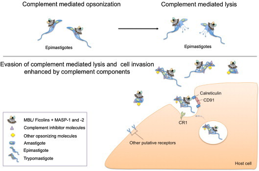
    1. Ambrosio A.R., De Messias-Reason I.J.T. Leishmania (Viannia) braziliensis: interaction of mannose-binding lectin with surface glycoconjugates and complement activation. An antibody-independent defence mechanism. Parasite Immunol. 2005;27:333–340. doi: 10.1111/j.1365-3024.2005.00782.x. - DOI - PubMed

Publication types

MeSH terms

Substances