Clinical Trial
. 2015 Jun 4;125(23):3637-46.
doi: 10.1182/blood-2015-02-629683. Epub 2015 Apr 10.

Modified Ham test for atypical hemolytic uremic syndrome

Affiliations
Clinical Trial

Modified Ham test for atypical hemolytic uremic syndrome

Eleni Gavriilaki et al. Blood. .

Abstract

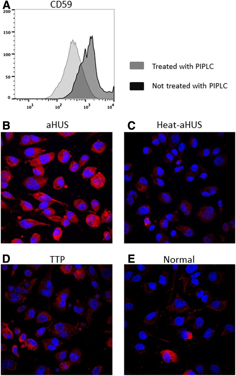
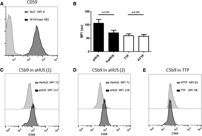
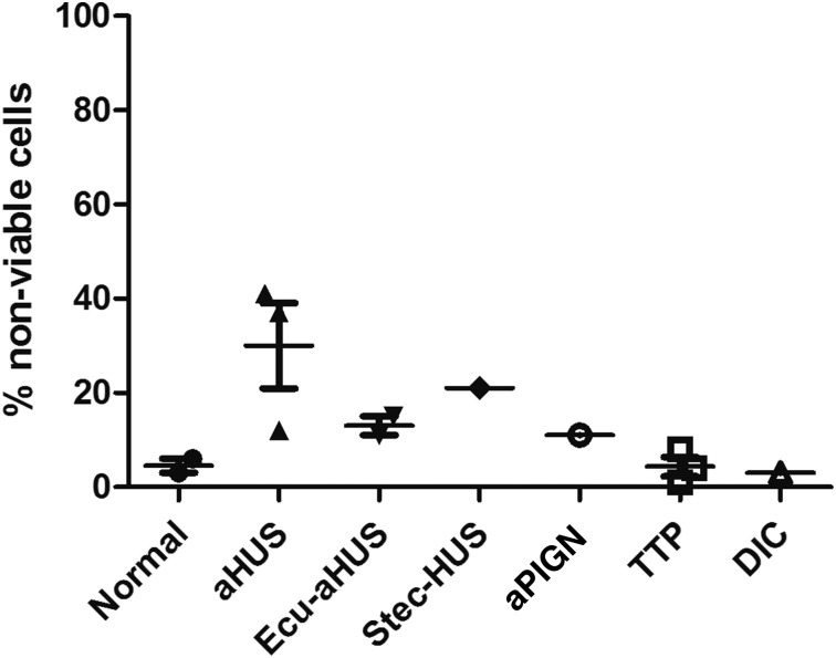
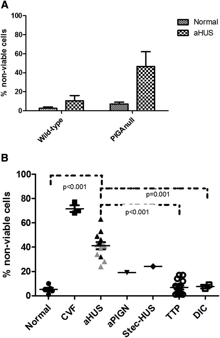
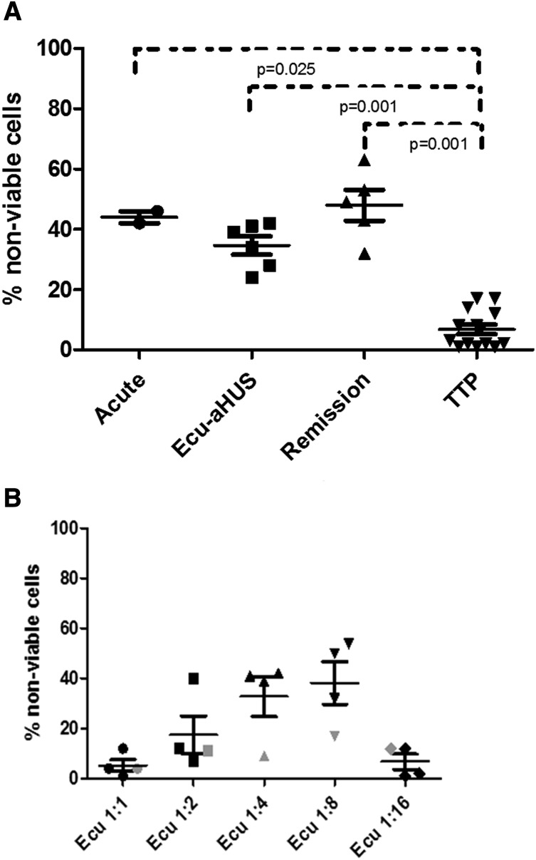
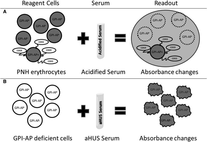
Atypical hemolytic uremic syndrome (aHUS) is a thrombotic microangiopathy (TMA) characterized by excessive activation of the alternative pathway of complement (APC). Atypical HUS is frequently a diagnosis of exclusion. Differentiating aHUS from other TMAs, especially thrombotic thrombocytopenic purpura (TTP), is difficult due to overlapping clinical manifestations. We sought to develop a novel assay to distinguish aHUS from other TMAs based on the hypothesis that paroxysmal nocturnal hemoglobinuria cells are more sensitive to APC-activated serum due to deficiency of glycosylphosphatidylinositol- anchored complement regulatory proteins (GPI-AP). Here, we demonstrate that phosphatidylinositol-specific phospholipase C-treated EA.hy926 cells and PIGA-mutant TF-1 cells are more susceptible to serum from aHUS patients than parental EA.hy926 and TF-1 cells. We next studied 31 samples from 25 patients with TMAs, including 9 with aHUS and 12 with TTP. Increased C5b-9 deposition was evident by confocal microscopy and flow cytometry on GPI-AP-deficient cells incubated with aHUS serum compared with heat-inactivated control, TTP, and normal serum. Differences in cell viability were observed in biochemically GPI-AP-deficient cells and were further increased in PIGA-deficient cells. Serum from patients with aHUS resulted in a significant increase of nonviable PIGA-deficient TF-1 cells compared with serum from healthy controls (P < .001) and other TMAs (P < .001). The cell viability assay showed high reproducibility, sensitivity, and specificity in detecting aHUS. In conclusion, we developed a simple, rapid, and serum-based assay that helps to differentiate aHUS from other TMAs.


Figures

Figure 1
Figure 1
C5b-9 deposition on PIPLC-treated human EA.hy926 cells. (A) Flow cytometric analysis of CD59 expression on ADP-activated EA.hy926 l cells treated with PIPLC vs untreated EA.hy926 cells. After trypsinization, endothelial cells were recovered in cell culture medium at 37°C for 30 minutes, treated with PIPLC, and then stained with anti-CD59 antibody. Reduced CD59 expression on PIPLC-treated cells is shown. (B-E) Confocal microscopy of ADP-activated EA.hy926 cells treated with PIPLC and stained with C5b-9 (depicted in red) and 4,6 diamidino-2-phenylindole (blue) as nuclei cell counterstaining. Magnification 40x. C5b-9 deposition is shown after incubation with aHUS serum (B) compared with heat-inactivated aHUS serum (C), TTP serum (D) and normal serum (E). PIPLC: Phosphatidylinositol-specific phospholipase C; aHUS: atypical hemolytic uremic syndrome; Heat-aHUS: heat inactivated atypical hemolytic uremic syndrome; TTP: thrombotic thrombocytopenic purpura.
Figure 2
Figure 2
C5b-9 deposition by flow cytometry on PIGA-null TF-1 cells. (A) Loss of CD59 expression on PIGA null TF-1 cells vs wild type TF-1 cells. (B) Comparison of C5b-9 deposition between aHUS and TTP samples and their heat-inactivated controls (HaHUS and HTTP, respectively). (C) Representative experiment of C5b-9 deposition after incubation with acute TTP serum (black) vs heat-inactivated control (light gray). (D) C5b-9 deposition after incubation with serum from a patient with aHUS in remission (black) compared with heat-inactivated control (light gray). (E) C5b-9 deposition after incubation with serum from a second patient with acute aHUS (black) compared with heat-inactivated control (light gray). HaHUS, heat-inactivated atypical hemolytic uremic syndrome serum; HTTP, heat-inactivated thrombotic thrombocytopenic purpura; MFI, mean fluorescence intensity.
Figure 3
Figure 3
WST-1 viability assay on PIPLC-treated EA.hy926 cells. Percentage of nonviable PIPLC-treated EA.hy926 cells among different disease entities (data are presented as mean with standard error of the mean [SEM]). aPIGN, atypical postinfectious glomerulonephritis; Ecu-aHUS, atypical hemolytic uremic syndrome treated with eculizumab; Stec-HUS: Shiga-toxin–associated hemolytic uremic syndrome.
Figure 4
Figure 4
Genetic disruption of PIGA augments cell killing in aHUS serum. (A) Comparison of nonviable cells between TF-1 wild-type and TF-1 PIGA-null cells (data are presented as mean with SEM) after incubation with normal and aHUS serum. (B) Percentage of nonviable PIGA-null TF1 cells among different disease entities (data are presented as mean with SEM). Serum from aHUS patients induces significantly increased percentages of nonviable cells compared with healthy controls (P < .001), TTP (P < .001), and DIC (P = .001). Gray triangles symbolize values of aHUS patients treated with eculizumab, whereas black triangles symbolize aHUS in acute phase or remission. CVF, CVF-activated serum; Stec-HUS, Shiga-toxin–associated hemolytic uremic syndrome.
Figure 5
Figure 5
WST-1 cell viability assay in patients with aHUS. (A) Similarly increased percentages of nonviable TF1 cells among aHUS patients in different disease states (acute phase, eculizumab treatment, and remission) compared with TTP patients. (B) Effects of serial dilution of eculizumab-treated serum on cell viability (1:1, 1:2, 1:4, 1:8, and 1:16). All aHUS patients showed no increase in killing at 1:1 and 1:16 dilutions, 2 of the 3 aHUS patients had no increase in killing at 1:2, and all 3 aHUS patients showed increased killing at 1:4 and 1:8 dilutions. Serum from a PNH patient treated with eculizumab (indicated by gray symbols) showed no increase in killing at any dilution. Ecu-aHUS, aHUS patient treated with eculizumab.
Figure 6
Figure 6
Model of Ham test for PNH and modified Ham test for aHUS diagnosis. The Ham test evaluates the effect of acidified serum on patient's cells (A), whereas the modified test evaluates the effect of patient's serum on GPI-AP–deficient reagent cells (B). Both tests use absorbance changes as readouts of cell viability.

Comment in

References

    1. Cataland SR, Wu HM. Diagnosis and management of complement mediated thrombotic microangiopathies. Blood Rev. 2014;28(2):67–74. - PubMed
    1. Nester CM, Thomas CP. Atypical hemolytic uremic syndrome: what is it, how is it diagnosed, and how is it treated? Hematology Am Soc Hematol Educ Program. 2012;2012:617-625. - PubMed
    1. Rennard S, Abe S. Decreased cold-insoluble globulin in congenital thrombocytopenia (Upshaw-Schulman syndrome). N Engl J Med. 1979;300(7):368. - PubMed
    1. Kinoshita S, Yoshioka A, Park YD, et al. Upshaw-Schulman syndrome revisited: a concept of congenital thrombotic thrombocytopenic purpura. Int J Hematol. 2001;74(1):101-108. - PubMed
    1. Furlan M, Robles R, Solenthaler M, Wassmer M, Sandoz P, Lammle B. Deficient activity of von Willebrand factor-cleaving protease in chronic relapsing thrombotic thrombocytopenic purpura. Blood. 1997;89(9):3097-3103. - PubMed

Publication types

Substances