. 2015 Jul;18(3):265-81.
doi: 10.1007/s10456-015-9465-6. Epub 2015 Apr 11.

Cardiovascular risk factors cause premature rarefaction of the collateral circulation and greater ischemic tissue injury

Affiliations

Cardiovascular risk factors cause premature rarefaction of the collateral circulation and greater ischemic tissue injury

Scott M Moore et al. Angiogenesis. 2015 Jul.

Abstract

Rationale: Collaterals lessen tissue injury in occlusive disease. However, aging causes progressive decline in their number and smaller diameters in those that remain (collateral rarefaction), beginning at 16 months of age in mice (i.e., middle age), and worse ischemic injury-effects that are accelerated in even 3-month-old eNOS(-/-) mice. These findings have found indirect support in recent human studies.

Objective: We sought to determine whether other cardiovascular risk factors (CVRFs) associated with endothelial dysfunction cause collateral rarefaction, investigate possible mechanisms, and test strategies for prevention.

Methods and results: Mice with nine different models of CVRFs of 4-12 months of age were assessed for number and diameter of native collaterals in skeletal muscle and brain and for collateral-dependent perfusion and ischemic injury after arterial occlusion. Hypertension caused collateral rarefaction whose severity increased with duration and level of hypertension, accompanied by greater hindlimb ischemia and cerebral infarct volume. Chronic treatment of wild-type mice with L-N (G)-nitro-arginine methylester caused similar rarefaction and worse ischemic injury which were not prevented by lowering arterial pressure with hydralazine. Metabolic syndrome, hypercholesterolemia, diabetes mellitus, and obesity also caused collateral rarefaction. Neither chronic statin treatment nor exercise training lessened hypertension-induced rarefaction.

Conclusion: Chronic CVRF presence caused collateral rarefaction and worse ischemic injury, even at relatively young ages. Rarefaction was associated with increased proliferation rate of collateral endothelial cells, effects that may promote accelerated endothelial cell senescence.


Conflict of interest statement

Disclosure/Conflict of Interest: None for any of the authors

Figures

Figure 1
Figure 1. Mean arterial pressure (MAP, via carotid artery, isoflurane anesthesia) and heart rate in wildtype littermates (WT) and renin transgenic (RTG) heterozygous (+/−) and homozygous (+/+) mice at the indicated ages (mos, months)
Values are mean ± SEM, number of animals (n) indicated in columns, *,**,*** p<0.05, 0.01, 0.001 levels of significance in this and subsequent figures.
Figure 2
Figure 2. Hypertension causes greater ischemia and impaired recovery of perfusion after femoral artery ligation (FAL)
A, Laser Doppler imaging (LDI) of plantar perfusion (dotted lines identify region of interest in representative figure), which correlates with whole limb perfusion, is reduced in 8 months-old RTG+/− mice immediately after FAL (post-op; inset bar graph), indicating reduced native collateral conductance. Lower recovery of perfusion on subsequent days is consistent with reduced collateral remodeling (confirmed in Figure 5). B, Perfusion in RTG+/− adductor region, which contains the major collateral network recruited by FAL (note flow induced after FAL in the two parallel-running collateral pathways in the gracilis muscles), is lower immediately after FAL (inset) and trends higher after day-7, consistent with the conclusions above for panel A. C, Ischemic nail-beds in RTG+/− mouse one day after FAL (arrows in representative figure). Hindlimb use-impairment and ischemic appearance scores are greater in RTG+/− mice. D–F, Similar results were obtained in 8 months-old C57BL/6 mice treated with L-NAME (LN) for 6.5 months and having similar hypertension as in RTG+/− mice (20 mmHg increase, Online Table 1). Concomitant administration of hydralazine (Hyd) normalizes the hypertension (Online Table 1), yet the perfusion deficit persists, suggesting that nitric oxide synthase inhibition rather than hypertension is the primary cause of deficient collateral conductance in L-NAME treated mice.
Figure 3
Figure 3. Chronic hypertension causes collateral rarefaction in skeletal muscle and brain
A, B, mouse groups shown in Figure 2. Eight months-old RTG+/− mice have fewer native collaterals (asterisks) of smaller diameter in abdominal wall skeletal muscle and brain (decreases shown at top of bars are relative to WT, here and in subsequent figures). C, D, Similar effects in L-NAME treated mice (LN) are not prevented by concomitant treatment with hydralazine (H). NS, non-significant.
Figure 4
Figure 4. Collateral rarefaction increases with duration and severity of hypertension
A, B, RTG+/− mice are born with the same number of native collaterals as WT (data not shown). By 4 months-age, collateral diameter is reduced; reduction of both number and diameter is evident with longer duration of hypertension. RTG+/+ mice with more severe hypertension evidence earlier collateral rarefaction. C, Consistent with collateral rarefaction shown in Figure 3 in skeletal muscle, plantar perfusion (by LDI) immediately after FAL in 4 months-old mice is inversely related to the severity of hypertension.
Figure 5
Figure 5. Larger infarct volume and less outward remodeling of collaterals evident 3 days after permanent middle cerebral artery occlusion in 8 months-old RTG+/− hypertensive mice
Remodeling is given as percent increase from baseline for collaterals between MCA and ACA trees (shown in representative figure).
Figure 6
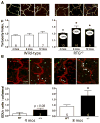
Figure 6. Tortuosity and proliferation in vascular wall cells are greater in native collaterals of hypertensive mice
Tortuosity (A) and proliferation of endothelial cells (B, arrows, EdU+ cells) in collaterals between MCA and ACA trees are increased during times shown in Figure 4 when rarefaction is occurring in RTG hypertensive mice.
Figure 7
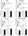
Figure 7. Cardiovascular risk factors in addition to hypertension cause rarefaction of the native collateral circulation
A, Eight months-old obese mice (Ob/Ob) have no deficit in hindlimb perfusion immediately after FAL (post-op, inset) or recovery of perfusion over subsequent 21 days, but have decreased collateral diameter. B, Eight months-old hyperlipidemic mice (LDLR−/−) have lower perfusion immediately after FAL and reduced recovery, and trend toward fewer collaterals. C, Eight months-old mice with Type 1 diabetes mellitus (DM, Akita) have lower perfusion immediately after FAL, impaired recovery, and trend towards fewer collaterals (see Online figure 4 and its legend showing significant rarefaction in a different model of Type 1 diabetes). D, Eight months-old mice with metabolic syndrome (2KO) have lower perfusion immediately after FAL, severe impairment in recovery of perfusion, and greater hindlimb use-impairment and ischemic appearance (Online Figure 5), and reduced collateral number and diameter.

References

    1. Go AS, Mozaffarian D, Roger VL, Benjamin EJ, Berry JD, Borden WB, Bravata DM, Dai S, Ford ES, Fox CS, Franco S, Fullerton HJ, Gillespie C, Hailpern SM, Heit JA, Howard VJ, Huffman MD, Kissela BM, Kittner SJ, Lackland DT, Lichtman JH, Lisabeth LD, Magid D, Marcus GM, Marelli A, Matchar DB, McGuire DK, Mohler ER, Moy CS, Mussolino ME, Nichol G, Paynter NP, Schreiner PJ, Sorlie PD, Stein J, Turan TN, Virani SS, Wong ND, Woo D, Turner MB. Executive summary: Heart disease and stroke statistics--2013 update: A report from the american heart association. Circulation. 2013;127:143–152. - PubMed
    1. Chin CT, Chen AY, Wang TY, Alexander KP, Mathews R, Rumsfeld JS, Cannon CP, Fonarow GC, Peterson ED, Roe MT. Risk adjustment for in-hospital mortality of contemporary patients with acute myocardial infarction: The acute coronary treatment and intervention outcomes network (action) registry-get with the guidelines (gwtg) acute myocardial infarction mortality model and risk score. Am Heart J. 2011;161:113–122. - PubMed
    1. Koennecke HC, Belz W, Berfelde D, Endres M, Fitzek S, Hamilton F, Kreitsch P, Mackert BM, Nabavi DG, Nolte CH, Pohls W, Schmehl I, Schmitz B, von Brevern M, Walter G, Heuschmann PU. Factors influencing in-hospital mortality and morbidity in patients treated on a stroke unit. Neurology. 2011;77:965–972. - PubMed
    1. Virkkunen J, Heikkinen M, Lepantalo M, Metsanoja R, Salenius JP. Diabetes as an independent risk factor for early postoperative complications in critical limb ischemia. J Vasc Surg. 2004;40:761–767. - PubMed
    1. Williams B, Menon M, Satran D, Hayward D, Hodges JS, Burke MN, Johnson RK, Poulose AK, Traverse JH, Henry TD. Patients with coronary artery disease not amenable to traditional revascularization: Prevalence and 3-year mortality. Catheter Cardiovasc Interv. 2010;75:886–891. - PubMed

Publication types

MeSH terms

LinkOut - more resources