. 2015 Jul;100(7):945-54.
doi: 10.3324/haematol.2014.122069. Epub 2015 Apr 10.

Targeting the spliceosome in chronic lymphocytic leukemia with the macrolides FD-895 and pladienolide-B

Affiliations

Targeting the spliceosome in chronic lymphocytic leukemia with the macrolides FD-895 and pladienolide-B

Manoj K Kashyap et al. Haematologica. 2015 Jul.

Abstract

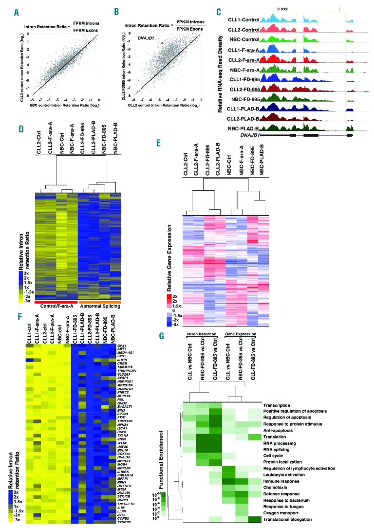
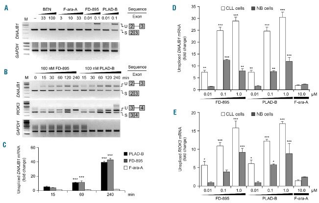
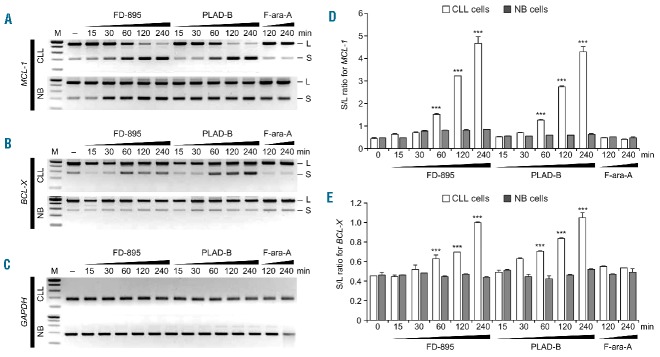
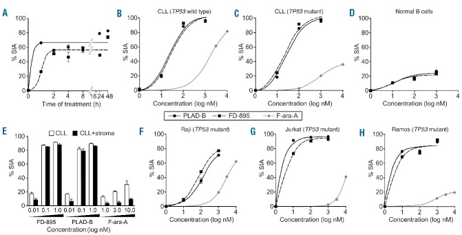
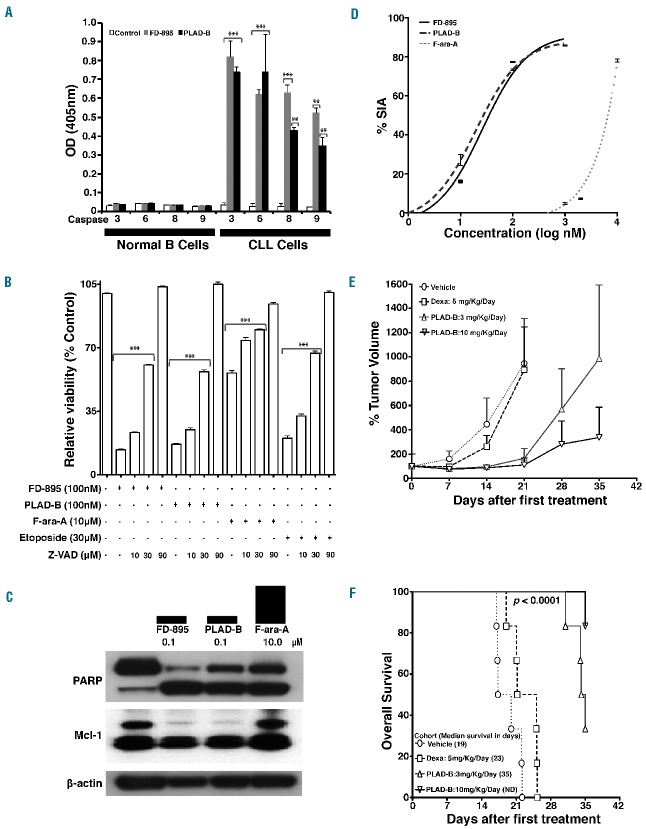
RNA splicing plays a fundamental role in human biology. Its relevance in cancer is rapidly emerging as demonstrated by spliceosome mutations that determine the prognosis of patients with hematologic malignancies. We report studies using FD-895 and pladienolide-B in primary leukemia cells derived from patients with chronic lymphocytic leukemia and leukemia-lymphoma cell lines. We found that FD-895 and pladienolide-B induce an early pattern of mRNA intron retention - spliceosome modulation. This process was associated with apoptosis preferentially in cancer cells as compared to normal lymphocytes. The pro-apoptotic activity of these compounds was observed regardless of poor prognostic factors such as Del(17p), TP53 or SF3B1 mutations and was able to overcome the protective effect of culture conditions that resemble the tumor microenvironment. In addition, the activity of these compounds was observed not only in vitro but also in vivo using the A20 lymphoma murine model. Overall, these findings give evidence for the first time that spliceosome modulation is a valid target in chronic lymphocytic leukemia and provide an additional rationale for the development of spliceosome modulators for cancer therapy.


Figures

Figure 1.
Figure 1.
RNA sequencing analysis in samples treated with FD-895 and PLAD-B. RNA transcriptome analysis was conducted on RNA obtained from two separate CLL samples (wild-type for TP53 and SF3B1) and normal B cells from healthy controls. CLL and normal B cells were incubated with 100 nM FD-895, 100 nM PLAD-B or 10 μM F-ara-A for 2 h prior to harvesting. (A) Comparison of IR log2 ratios [fragments per kilobase per million fragments mapped (FPKM) intron / FPKM exon] in untreated CLL and untreated normal B cells. The black line represents the diagonal where IR ratios are equal in both samples. (B) Comparison of IR log2 ratios in CLL control and FD-895-treated samples. (C) RNA-seq read densities at the DNAJB1 locus, a sample gene with high IR pattern. FD-895- and PLAD-B-treated samples but not F-ara-A-treated or untreated controls showed a pattern of IR with accumulation of read densities in the regions expanding introns 1 and 2. (D) Hierarchical clustering for approximately 3,500 genes shown in the heat-map depicting the relative IR log2 ratios. (E) Gene expression across normal B cells and CLL samples treated with FD-895, PLAD-B and F-ara-A shown in the heat-map for approximately 4,000 genes. A similar clustering was observed with additional CLL and normal B-cell samples. (F) RNA-seq heat map showing 50 genes with a high level of IR after treatment with either FD-895 or PLAD-B. (G) Heat-map showing gene ontology pathway enrichment (P-values) using genes with greater than 3-fold increases in either their IR or mRNA expression ratios in CLL or normal B cells in untreated control samples compared with samples incubated with FD-895.
Figure 2.
Figure 2.
Induction of intron retention / spliceosome modulation mediated by FD-895 and PLAD-B. (A) RT-PCR was used to access spliced (S) and unspliced (U) isoforms of DNAJB1 and GAPDH in CLL cells. (B) IR was evaluated by RT-PCR for DNAJB1, RIOK3 and GAPDH (used as loading control) in CLL cells after treatment. (C) Quantitative real-time-PCR analysis of CLL cells after treatment to evaluate levels of unspliced mRNA for DNAJB1. GAPDH was used for normalization. (D) Intron retention for DNAJB1 and (E) RIOK3 was evaluated by quantitative real-time-PCR in CLL and normal B cells using specific primers that allowed detection of intron-containing regions (Online Supplementary Table S1). The untreated controls (CLL cells or NBC) for DNAJB1 and RIOK3 were set to a value of 1. GAPDH was used as a control for normalization.
Figure 3.
Figure 3.
Regulation of alternative splicing after treatment with FD-895 or PLAD-B. (A–C) Normal B cells and CLL cells were treated with 100 nM FD-895, 100 nM PLAD-B or 10 μM F-ara-A for the indicated times. RT-PCR for (A) MCL-1, (B) BCL-X and (C) GAPDH was conducted. The RT-PCR generated two different products; a long or anti-apoptotic isoform (L) and a short or pro-apoptotic splice isoform (S). (D–E) The levels of the L and S isoforms were evaluated after incubation of 100 nM FD-895, 100 nM PLAD-B or 10 μM F-ara-A by densitometry of RT-PCR bands stained with ethidium bromide using Quantity One software (Bio-Rad). The S/L ratio was plotted as a mean of two separate experiments as shown in panel (D) for MCL-1 and (E) for BCL-X. Error bars represent SD and ***P<0.001.
Figure 4.
Figure 4.
FD-895 or PLAD-B induced apoptosis in CLL cells but not in normal lymphocytes with TP53-independent activity. (A) Primary leukemia B cells alone from a CLL patient were treated with 100 nM FD-895 or 100 nM PLAD-B at time 0 and allowed to incubate for 0.5 h, to 48 h. After initial incubations, the cells were washed twice with fresh media to remove the remaining FD-895 or PLAD-B and then were cultured using media only for a total of 48 h. The dark black line indicates FD-895 and the light color line indicates PLAD-B. Apoptosis was measured by flow cytometry using a PI/DiOC6 assay. (B–D) Cells from different sources were evaluated for their level of apoptosis after incubation with FD-895, PLAD-B or F-ara-A for 48 h, including: (B) cells from a CLL patient with wild-type TP53 and sensitive to F-ara-A; (C) cells from a CLL patient with Del(17p) and TP53 mutation (TP53-mut), resistant to F-ara-A; and, (D) normal B cells from a healthy donor. (E) Primary CLL cells derived from a Del(17p) patient were treated alone or co-cultured with stroma-NK-tert cells with FD-895, PLAD-B, or F-ara-A for 48 h. Apoptosis was measured by flow cytometry using a CD19/CD5/DiOC6 assay. (F-H) In parallel, cell lines with different TP53 mutation status were incubated with FD-895, PLAD-B or F-ara-A: (F) Raji - TP53 mutant type; (G) Jurkat cells - TP53 mutant; (H) Ramos cells - TP53 mutant. After incubation, apoptosis was assessed by flow cytometry. These results are representative of at least three separate experiments. In order to discriminate the compound specific-induced apoptosis vs. background spontaneous cell death from in vitro culture conditions, we calculated the percentage of specific induced apoptosis (% SIA) using the following formula: % SIA = [(compound induced apoptosis − media only spontaneous apoptosis) / (100− media only spontaneous apoptosis)] × 100.
Figure 5.
Figure 5.
FD-895 and PLAD-B induce apoptosis in CLL cells independent of TP53 and SF3B1 mutational status. Primary leukemia B cells were obtained from CLL patients with wild-type SF3B1 (n=10), SF3B1 mutant (n=10), or Del(17p)/TP53 mutations (n=10). Normal B cells (n=10) and T lymphocytes (n=10) were obtained from healthy donors. Samples were independently incubated with (A) 100 nM FD-895 or (B) 100 nM PLAD-B for 48 h. After incubation, the samples were harvested and percentage of specific induced apoptosis (% SIA) was measured by flow cytometry. FD-895 and PLAD-B induced apoptosis in CLL cells regardless of Del(17p)/TP53 or SF3B1 mutation status, while normal B cells or T cells showed statistically-lower levels of apoptosis. The data show the results of samples analyzed in duplicate with the means and their respective SD.
Figure 6.
Figure 6.
Effects of FD-895 and PLAD-B on modulation of caspases and apoptosis-associated proteins and in vivo effect on the A20 murine lymphoma BALB/c model. (A) CLL cells and normal B cells were assessed for caspase activation after treatment. Cells incubated with only media served as the control. (B) CLL cells were incubated with 100 nM FD-895, 100 nM PLAD-B, 10 μM F-ara-A, or 30 μM etoposide for 48 h either alone or in combination with different concentrations of Z-VAD (10 μM, 30 μM, or 90 μM). Following incubations, flow cytometry was performed using CD19/CD5/annexin-V to assess cell death. (C) CLL cells were incubated with 100 nM FD-895 or 100 nM PLAD-B for 6 h. Protein lysates from treated samples were analyzed by western blot using antibodies against PARP and Mcl-1. Cells incubated in media only were used as the negative control (−). (D) Induction of apoptosis in A20 cells after 48 h of treatment. (E) Tumor-bearing mice received intraperitoneal injections consecutively for 5 days with either vehicle, dexamethasone, PLAD-B low or high dose (3 mg/kg/day or 10 mg/kg/day). Tumor volume was measured over time. (F) Survival of mice treated as described above and followed for 35 days after injection; the error bars indicate SD. Survival was calculated using the Kaplan–Meier method.

References

    1. Rai KR. Pathophysiologic mechanisms of chronic lymphocytic leukemia and their application to therapy. Exp Hematol. 2007;35(4 Suppl 1):134–136. - PubMed
    1. Hallek M, Fischer K, Fingerle-Rowson G, et al. Addition of rituximab to fludarabine and cyclophosphamide in patients with chronic lymphocytic leukaemia: a randomised, open-label, phase 3 trial. Lancet. 2010;376(9747):1164–1174. - PubMed
    1. Castro JE. Treatment of patients with chronic lymphocytic leukemia with 17p deletion: the saga continues. Leuk Lymphoma. 2012;53(2):179–180. - PubMed
    1. Hallek M. Signaling the end of chronic lymphocytic leukemia: new frontline treatment strategies. Blood. 2013;122(23):3723–3734. - PubMed
    1. Kornblihtt AR, Schor IE, Alló M, Dujardin G, Petrillo E, Muñoz MJ. Alternative splicing: a pivotal step between eukaryotic transcription and translation. Nat Rev Mol Cell Biol. 2013;14(3):153–165. - PubMed

Publication types

MeSH terms