

Oxidative stress and Treg depletion in lupus patients with anti-phospholipid syndrome
- PMID: 25862984
- PMCID: PMC4464983
- DOI: 10.1016/j.clim.2015.03.024
Oxidative stress and Treg depletion in lupus patients with anti-phospholipid syndrome
Abstract
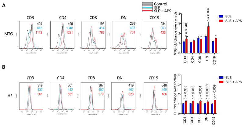
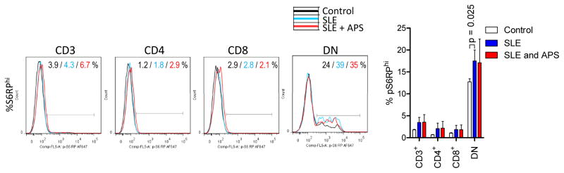
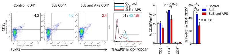
Anti-phospholipid antibodies (APLA) represent a diagnostic criterion of systemic lupus erythematosus (SLE) and cause morbidity, termed anti-phospholipid syndrome (APS). Activation of the mechanistic target of rapamycin (mTOR) has been recently associated with APS. mTOR is a sensor of oxidative stress. Therefore, we examined mitochondrial mass, superoxide production, mTOR activity and FoxP3 expression in 72 SLE patients, twelve of whom also had APS, and 54 healthy controls by flow cytometry. Mitochondrial mass was increased in CD4(-)CD8(-) double-negative (DN) T cells of SLE patients with APS (2.7-fold) in comparison to those without APS (1.7-fold; p = 0.014). Superoxide production was increased in all lymphocyte subsets of APS patients. FoxP3(+) cells were depleted within CD4(+)CD25(+) Tregs in patients with APS (28.4%) relative to those without APS (46.3%, p = 0.008). mTOR activity was similar between SLE patients with and without APS. Thus, oxidative stress and Treg depletion rather than mTOR activation underlie APS in patients with SLE.
Keywords: Anti-phospholipid syndrome; Mechanistic target of rapamycin; Oxidative stress; Systemic lupus erythematosus; Treg; mTOR.
Copyright © 2015 Elsevier Inc. All rights reserved.
Figures
References
-
- Tsokos GC. Systemic Lupus Erythematosus. N Engl J Med. 2011;365:2110–2121. - PubMed
-
- Lai Z-W, Borsuk R, Shadakshari A, Yu J, Dawood M, Garcia R, Francis L, Tily H, Bartos A, Faraone SV, Phillips PE, Perl A. mTOR activation triggers IL-4 production and necrotic death of double-negative T cells in patients with systemic lupus eryhthematosus. J Immunol. 2013;191:2236–2246. - PMC - PubMed
-
- Lai ZW, Hanczko R, Bonilla E, Caza TN, Clair B, Bartos A, Miklossy G, Jimah J, Doherty E, Tily H, Francis L, Garcia R, Dawood M, Yu J, Ramos I, Coman I, Faraone SV, Phillips PE, Perl A. N-acetylcysteine reduces disease activity by blocking mTOR in T cells of lupus patients. Arthritis Rheum. 2012;64:2937–2946. - PMC - PubMed
Publication types
MeSH terms
Substances
Grants and funding
LinkOut - more resources
Full Text Sources
Other Literature Sources
Medical
Research Materials
Miscellaneous
