

Altered mechanical environment of bone cells in an animal model of short- and long-term osteoporosis
- PMID: 25863050
- PMCID: PMC4390805
- DOI: 10.1016/j.bpj.2015.02.031
Altered mechanical environment of bone cells in an animal model of short- and long-term osteoporosis
Abstract
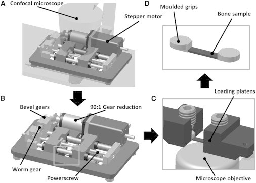
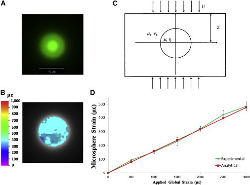
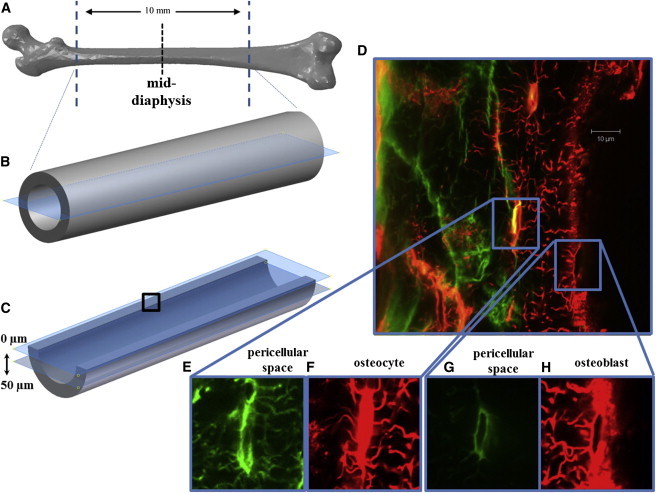
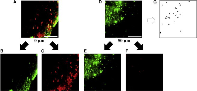
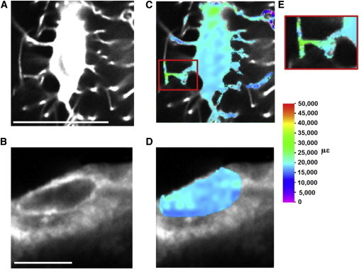
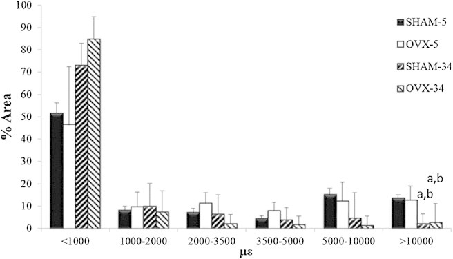
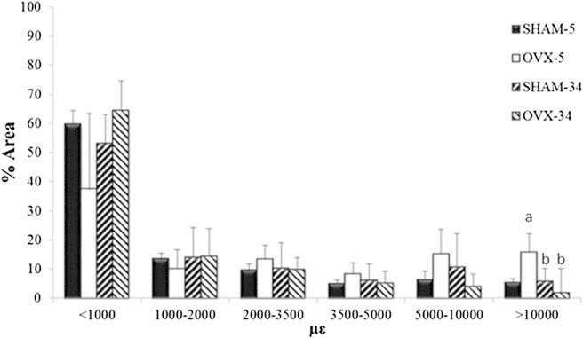
Alterations in bone tissue composition during osteoporosis likely disrupt the mechanical environment of bone cells and may thereby initiate a mechanobiological response. It has proved challenging to characterize the mechanical environment of bone cells in vivo, and the mechanical environment of osteoporotic bone cells is not known. The objective of this research is to characterize the local mechanical environment of osteocytes and osteoblasts from healthy and osteoporotic bone in a rat model of osteoporosis. Using a custom-designed micromechanical loading device, we apply strains representative of a range of physical activity (up to 3000 με) to fluorescently stained femur samples from normal and ovariectomized rats. Confocal imaging was simultaneously performed, and digital image correlation techniques were applied to characterize cellular strains. In healthy bone tissue, osteocytes experience higher maximum strains (31,028 ± 4213 με) than osteoblasts (24,921 ± 3,832 με), whereas a larger proportion of the osteoblast experiences strains >10,000 με. Most interestingly, we show that osteoporotic bone cells experience similar or higher maximum strains than healthy bone cells after short durations of estrogen deficiency (5 weeks), and exceeded the osteogenic strain threshold (10,000 με) in a similar or significantly larger proportion of the cell (osteoblast, 12.68% vs. 13.68%; osteocyte, 15.74% vs. 5.37%). However, in long-term estrogen deficiency (34 weeks), there was no significant difference between bone cells in healthy and osteoporotic bone. These results suggest that the mechanical environment of bone cells is altered during early-stage osteoporosis, and that mechanobiological responses act to restore the mechanical environment of the bone tissue after it has been perturbed by ovariectomy.
Copyright © 2015 Biophysical Society. Published by Elsevier Inc. All rights reserved.
Figures
Comment in
-
Seeing the unseen: cell strain and mechanosensing.Biophys J. 2015 Apr 7;108(7):1583-1584. doi: 10.1016/j.bpj.2015.03.008. Biophys J. 2015. PMID: 25863048 Free PMC article. No abstract available.
References
-
- Birmingham E., Niebur G.L., McNamara L.M. Osteogenic differentiation of mesenchymal stem cells is regulated by osteocyte and osteoblast cells in a simplified bone niche. Eur. Cell. Mater. 2012;23:13–27. - PubMed
-
- Jahani M., Genever P.G., Fagan M.J. The effect of osteocyte apoptosis on signaling in the osteocyte and bone lining cell network: a computer simulation. J. Biomech. 2012;45:2876–2883. - PubMed
-
- Klein-Nulend J., van der Plas A., Burger E.H. Sensitivity of osteocytes to biomechanical stress in vitro. FASEB J. 1995;9:441–445. - PubMed
-
- Owan I., Burr D.B., Duncan R.L. Mechanotransduction in bone: osteoblasts are more responsive to fluid forces than mechanical strain. Am. J. Physiol. Cell Physiol. 1997;273:C810–C815. - PubMed
-
- Smalt R., Mitchell F.T., Chambers T.J. Induction of NO and prostaglandin E2 in osteoblasts by wall-shear stress but not mechanical strain. Am. J. Physiol. Endocrinol. Metab. 1997;273:E751–E758. - PubMed
Publication types
MeSH terms
Substances
LinkOut - more resources
Full Text Sources
Other Literature Sources
Medical
