. 2015 Apr 7;108(7):1599-1603.
doi: 10.1016/j.bpj.2015.02.007.

Front-to-rear membrane tension gradient in rapidly moving cells

Affiliations

Front-to-rear membrane tension gradient in rapidly moving cells

Arnon D Lieber et al. Biophys J. .

Abstract

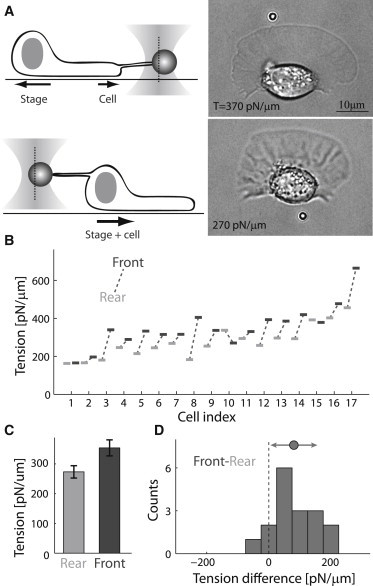
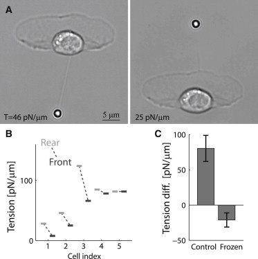
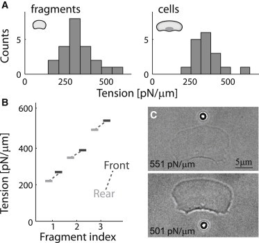
Membrane tension is becoming recognized as an important mechanical regulator of motile cell behavior. Although membrane-tension measurements have been performed in various cell types, the tension distribution along the plasma membrane of motile cells has been largely unexplored. Here, we present an experimental study of the distribution of tension in the plasma membrane of rapidly moving fish epithelial keratocytes. We find that during steady movement the apparent membrane tension is ∼30% higher at the leading edge than at the trailing edge. Similar tension differences between the front and the rear of the cell are found in keratocyte fragments that lack a cell body. This front-to-rear tension variation likely reflects a tension gradient developed in the plasma membrane along the direction of movement due to viscous friction between the membrane and the cytoskeleton-attached protein anchors embedded in the membrane matrix. Theoretical modeling allows us to estimate the area density of these membrane anchors. Overall, our results indicate that even though membrane tension equilibrates rapidly and mechanically couples local boundary dynamics over cellular scales, steady-state variations in tension can exist in the plasma membranes of moving cells.


Figures

Figure 1
Figure 1
Membrane tension is higher at the leading edge of motile keratocytes. (A) Schematic illustrations (left) and bright-field images (right) of a motile keratocyte during membrane-tension measurements at the front (upper) and rear (lower). The time interval between the front and rear tension measurements was 22 s, and the tension value at each position is indicated in the images. The focal planes of the images are slightly shifted due to differences in bead height when pulling from the front as compared to pulling from the rear. (B) Front (dark) and rear (light) membrane-tension values are shown for 17 different cells. (C) Bar plot showing the population-averaged membrane-tension values at the front and rear of the cell (mean ± SE). The difference in the average tension between front and rear is statistically significant (p < 0.01). (D) Histogram of the front-to-rear membrane-tension difference distribution for the population of cells shown in (B). The mean and standard deviation are indicated above the histogram.
Figure 2
Figure 2
Membrane tension in frozen cells is higher at the rear. (A) Bright-field images of a cell that has stopped moving after treatment with blebbistatin followed by jasplakinolide, during membrane tension measurements at the front and rear. (B) Front (dark) and rear (light) membrane-tension values are shown for different frozen cells. (C) Bar plot showing the population-averaged membrane-tension difference between the front and rear of the cell (mean ± SE) in control cells and frozen cells. In contrast with control cells, the tension values in frozen cells are higher at the rear. The difference in the average tension difference between control cells and frozen cells is statistically significant (p < 0.01).
Figure 3
Figure 3
Membrane tension gradient in lamellipodial fragments. (A) Histograms of membrane tension values at the leading edge in a population of lamellipodial fragments (left) and whole cells (right). The membrane tension in fragments is comparable to that in cells (p > 0.1). (B) Front (dark) and rear (light) tension values are shown for three fragments. (C) Bright-field images of a fragment during membrane-tension measurements at the front and rear. The time interval between the front and rear tension measurements was 80 s.

References

    1. Houk A.R., Jilkine A., Weiner O.D. Membrane tension maintains cell polarity by confining signals to the leading edge during neutrophil migration. Cell. 2012;148:175–188. - PMC - PubMed
    1. Gauthier N.C., Masters T.A., Sheetz M.P. Mechanical feedback between membrane tension and dynamics. Trends Cell Biol. 2012;22:527–535. - PubMed
    1. Lieber A.D., Yehudai-Resheff S., Keren K. Membrane tension in rapidly moving cells is determined by cytoskeletal forces. Curr. Biol. 2013;23:1409–1417. - PubMed
    1. Keren K. Cell motility: the integrating role of the plasma membrane. Eur. Biophys. J. 2011;40:1013–1027. - PMC - PubMed
    1. Gauthier N.C., Fardin M.A., Sheetz M.P. Temporary increase in plasma membrane tension coordinates the activation of exocytosis and contraction during cell spreading. Proc. Natl. Acad. Sci. USA. 2011;108:14467–14472. - PMC - PubMed

Publication types

Substances