

The effect of substrate stiffness, thickness, and cross-linking density on osteogenic cell behavior
- PMID: 25863052
- PMCID: PMC4390808
- DOI: 10.1016/j.bpj.2015.02.022
The effect of substrate stiffness, thickness, and cross-linking density on osteogenic cell behavior
Abstract
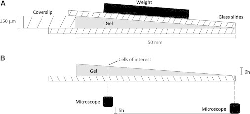
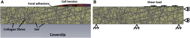
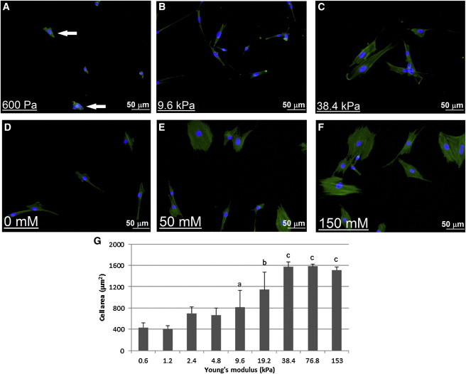
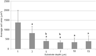
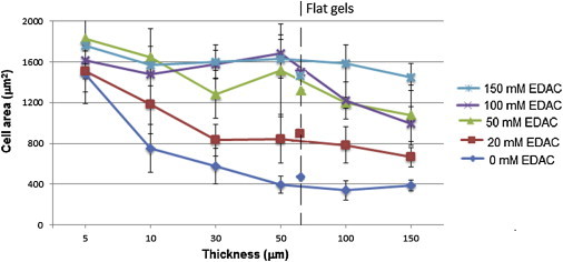
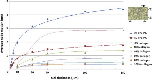
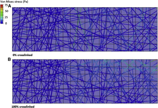
Osteogenic cells respond to mechanical changes in their environment by altering their spread area, morphology, and gene expression profile. In particular, the bulk modulus of the substrate, as well as its microstructure and thickness, can substantially alter the local stiffness experienced by the cell. Although bone tissue regeneration strategies involve culture of bone cells on various biomaterial scaffolds, which are often cross-linked to enhance their physical integrity, it is difficult to ascertain and compare the local stiffness experienced by cells cultured on different biomaterials. In this study, we seek to characterize the local stiffness at the cellular level for MC3T3-E1 cells plated on biomaterial substrates of varying modulus, thickness, and cross-linking concentration. Cells were cultured on flat and wedge-shaped gels made from polyacrylamide or cross-linked collagen. The cross-linking density of the collagen gels was varied to investigate the effect of fiber cross-linking in conjunction with substrate thickness. Cell spread area was used as a measure of osteogenic differentiation. Finite element simulations were used to examine the effects of fiber cross-linking and substrate thickness on the resistance of the gel to cellular forces, corresponding to the equivalent shear stiffness for the gel structure in the region directly surrounding the cell. The results of this study show that MC3T3 cells cultured on a soft fibrous substrate attain the same spread cell area as those cultured on a much higher modulus, but nonfibrous substrate. Finite element simulations predict that a dramatic increase in the equivalent shear stiffness of fibrous collagen gels occurs as cross-linking density is increased, with equivalent stiffness also increasing as gel thickness is decreased. These results provide an insight into the response of osteogenic cells to individual substrate parameters and have the potential to inform future bone tissue regeneration strategies that can optimize the equivalent stiffness experienced by a cell.
Copyright © 2015 Biophysical Society. Published by Elsevier Inc. All rights reserved.
Figures

References
-
- Tanaka-Kamioka K., Kamioka H., Lim S.-S. Osteocyte shape is dependent on actin filaments and osteocyte processes are unique actin-rich projections. J. Bone Miner. Res. 1998;13:1555–1568. - PubMed
-
- Kapur S., Baylink D.J., Lau K.H. Fluid flow shear stress stimulates human osteoblast proliferation and differentiation through multiple interacting and competing signal transduction pathways. Bone. 2003;32:241–251. - PubMed
-
- Engler A.J., Sen S., Discher D.E. Matrix elasticity directs stem cell lineage specification. Cell. 2006;126:677–689. - PubMed
MeSH terms
Substances
LinkOut - more resources
Full Text Sources
Other Literature Sources
