. 2015 Jun;185(6):1676-85.
doi: 10.1016/j.ajpath.2015.02.014. Epub 2015 Apr 8.

Heparanase stimulates chondrogenesis and is up-regulated in human ectopic cartilage: a mechanism possibly involved in hereditary multiple exostoses

Affiliations

Heparanase stimulates chondrogenesis and is up-regulated in human ectopic cartilage: a mechanism possibly involved in hereditary multiple exostoses

Julianne Huegel et al. Am J Pathol. 2015 Jun.

Abstract

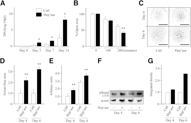
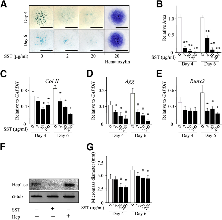
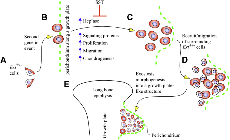
Hereditary multiple exostoses is a pediatric skeletal disorder characterized by benign cartilaginous tumors called exostoses that form next to growing skeletal elements. Hereditary multiple exostoses patients carry heterozygous mutations in the heparan sulfate (HS)-synthesizing enzymes EXT1 or EXT2, but studies suggest that EXT haploinsufficiency and ensuing partial HS deficiency are insufficient for exostosis formation. Searching for additional pathways, we analyzed presence and distribution of heparanase in human exostoses. Heparanase was readily detectable in most chondrocytes, particularly in cell clusters. In control growth plates from unaffected persons, however, heparanase was detectable only in hypertrophic zone. Treatment of mouse embryo limb mesenchymal micromass cultures with exogenous heparanase greatly stimulated chondrogenesis and bone morphogenetic protein signaling as revealed by Smad1/5/8 phosphorylation. It also stimulated cell migration and proliferation. Interfering with HS function both with the chemical antagonist Surfen or treatment with bacterial heparitinase up-regulated endogenous heparanase gene expression, suggesting a counterintuitive feedback mechanism that would result in further HS reduction and increased signaling. Thus, we tested a potent heparanase inhibitor (SST0001), which strongly inhibited chondrogenesis. Our data clearly indicate that heparanase is able to stimulate chondrogenesis, bone morphogenetic protein signaling, cell migration, and cell proliferation in chondrogenic cells. These properties may allow heparanase to play a role in exostosis genesis and pathogenesis, thus making it a conceivable therapeutic target in hereditary multiple exostoses.


Figures

Figure 1
Figure 1
Heparanase is broadly distributed in human exostoses but restricted in control growth plate. A–D: Immunohistochemical staining of longitudinal sections of control human growth plate shows that heparanase is detectable in hcs at the coj and is also prominent in perichondrium (C, arrowheads). Strong staining is appreciable in a blood vessel present in the SOC as expected (D). E–H: Sections from human exostoses were stained in parallel. Heparanase staining is clear in nearly every chondrocyte regardless of location within tissue and apparent phenotypic maturation status (E and F) and also in neighboring perichondrium-like tissue (G, arrowheads). Clusters of large hypertrophying chondrocytes display strong staining, particularly in their pericellular compartment (H). I: Continuous sections from the above exostosis specimen were stained with collagen X antibodies. Note that only mature and hcs stain positively. B and F are higher magnification images of areas in A and E, respectively. Scale bars: 250 μm (A and E); 75 μm (B, D, F, G, H, and I); 300 μm (C). Coj, chondro-osseous junction; hc, hypertrophic chondrocyte; pc, perichondrium; phc, pre-hypertrophic cartilage; rc/pl, resting-proliferating cartilage; SOC, secondary ossification center.
Figure 2
Figure 2
Treatment with human recombinant heparanase stimulates cell proliferation, migration, and chondrogenesis. A: DNA quantification shows that heparanase stimulates proliferation of ATDC5 cells in monolayer as early as 3 days after treatment compared with vehicle-treated controls. B: Scratch wound-healing assays show that cell migration is increased by heparanase treatment. Wound closure was monitored by sequential phase contrast imaging up to 3 hours. C: Representative images of stained limb bud cell micromass cultures on day 4 and 6 treated with recombinant heparanase or vehicle. Note the substantial increase in the number and staining intensity of cartilage nodules in treated versus control cultures. D: Image-based quantification of Alcian Blue–positive areas shows more than twofold increase in treated cultures over control set at 1. E: Measurement of overall micromass diameter on day 4 and 6 indicates that proliferation/migration of peripheral cells was stimulated by heparanase treatment. Increase in diameter for each micromass was set relative to that at 24 hours after plating and is described in arbitrary units. F: Immunoblot images show that heparanase treatment had increased Smad1/5/8 phosphorylation protein levels in day 4 and 6 micromass cultures relative to respective controls. Membranes were re-blotted with α-tub antibodies for sample loading normalization. G: Normalized band intensity quantifications of blots in F with controls set at 1. n = 3 (A); n = 6 (B, D, and E). P < 0.05, ∗∗P < 0.01. Scale bar = 1.5 mm. Cntl, control; Hep'ase, heparanase protein; α-tub, α-tubulin.
Figure 3
Figure 3
Heparanase expression is responsive to modulation in heparan sulfate levels or function. A–D: Immunoblot images and densitometric histograms indicate that endogenous heparanase protein levels increase by Surfen treatment (+) in limb bud micromasses (A and B) and ATDC5 cell cultures (C and D) compared with respective untreated controls (−). E and F: Semiquantitative RT-PCR and densitometric analyses show that Surfen treatment increases heparanase gene expression in micromass cultures. G and H: Immunoblot and densitometric quantification analyses show up-regulation of endogenous heparanase protein levels on treatment with recombinant BMP2. Data are expressed as means ± SD. P < 0.05. BMP, bone morphogenetic protein; Cntl, control; Hep'ase, heparanase protein; Surfen, bis-2-methyl-4-aminoquinolyl-6-carbamide; α-tub, α-tubulin.
Figure 4
Figure 4
Chondrogenesis is inhibited by a heparanase antagonist. A: Images of Alcian Blue–stained micromass cultures on day 4 and 6 show a dose-dependent reduction in cartilage nodule formation on treatment with the heparanase antagonist SST0001. Panels on the right show images of control and treated micromass cultures counterstained with hematoxylin. B: Image-assisted quantification of Alcian Blue–positive area in control versus SST0001-treated cultures. Statistical significance was reached at every concentration tested. C–E: Histograms depict gene expression levels of chondrogenic genes ColII, Agg, and Runx2 in controls versus SST0001-treaed micromass cultures on day 4 and 6 measured by semiquantitative RT-PCR and quantified by imaging. F: Immunoblot images show decrease in the endogenous Hep'ase levels in micromass cultures on SST0001 treatment and increase on bacterial Hep treatment compared with control. G: Histograms depict the overall absolute micromass diameter at day 4 and 6 in control versus SST0001-treated cultures. Data are expressed as means ± SD. n = 2 (CE); n = 6 (B and G). P < 0.05, ∗∗P < 0.01. Scale bar = 3 mm. Hep, heparitinase; Hep'ase, heparanase protein; SST, SST0001.
Figure 5
Figure 5
Model illustrates a series of regulatory steps that may cause inception and promotion of formation and growth of exostosis. A: The cells of most patients with hereditary multiple exostoses bear a heterozygous loss-of-function mutation in EXT1 or EXT2 (depicted here at Ext+/− cells in half white and half red). B: At one point prenatally or postnatally, some cells may undergo a second genetic change (depicted here as red cells along perichondrium and called mutant in the text), resulting in a more severe loss of overall EXT expression or function and leading to further reduction in local HS production and levels. C: This in turn may up-regulate Hep'ase expression, increase growth factor availability and cell proliferation, and induce ectopic chondrogenesis near/at perichondrium. D: The incipient exostosis cells may recruit surrounding heterozygous cells, induce them into neoplastic behavior, and promote their incorporation into the outgrowing exostosis. E: Cells within the outgrowing exostosis may assemble into a typical growth plate-like structure that protrudes away from the surface of the skeletal element (depicted here as a long bone) and cover distally by perichondrium. By inhibiting heparanase activity and possibly other processes, SST may inhibit chondrogenesis and in turn exostosis formation as indicated. Surfen (not indicated here) may elicit the opposite and stimulate chondrogenesis. Hep'ase, heparanase; HS, heparan sulfate; SST, SST0001; Surfen, bis-2-methyl-4-aminoquinolyl-6-carbamide.

References

    1. Fux L., Ilan N., Sanderson R.D., Vlodavsky I. Heparanase: busy at the cell surface. Trends Biochem Sci. 2009;34:511–519. - PMC - PubMed
    1. Vreys V., David G. Mammalian heparanase: what is the message? J Cell Mol Med. 2007;11:427–452. - PMC - PubMed
    1. Freeman C., Parish C.R. Human platelet heparanase: purification, characterization and catalytic activity. Biochem J. 1998;330:1341–1350. - PMC - PubMed
    1. Levy-Adam F., Feld S., Suss-Toby E., Vlodavsky I., Ilan N. Heparanase facilitates cell adhesion and spreading by clustering of cell surface heparan sulfate proteoglycans. PLoS ONE. 2008;3:e2319. - PMC - PubMed
    1. Patel V.N., Knox S.M., Likar K.M., Lathrop C.A., Hossain R., Eftekhari S., Whitelock J.M., Elkin M., Vlodavsky I., Hoffman M.P. Heparanase cleavage of perlecan heparan sulfate modulates FGF10 activity during ex vivo submandibular gland branching morphogenesis. Development. 2007;134:4177–4186. - PubMed

Publication types

MeSH terms