

Evidence for early neurodegeneration in the cervical cord of patients with primary progressive multiple sclerosis
- PMID: 25863355
- PMCID: PMC4614136
- DOI: 10.1093/brain/awv086
Evidence for early neurodegeneration in the cervical cord of patients with primary progressive multiple sclerosis
Abstract
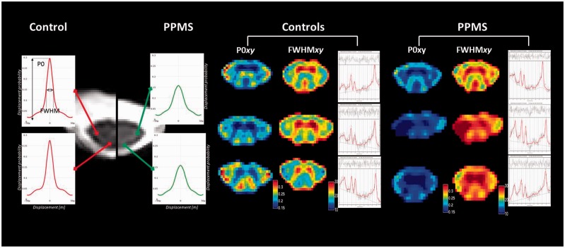
Spinal neurodegeneration is an important determinant of disability progression in patients with primary progressive multiple sclerosis. Advanced imaging techniques, such as single-voxel (1)H-magnetic resonance spectroscopy and q-space imaging, have increased pathological specificity for neurodegeneration, but are challenging to implement in the spinal cord and have yet to be applied in early primary progressive multiple sclerosis. By combining these imaging techniques with new clinical measures, which reflect spinal cord pathology more closely than conventional clinical tests, we explored the potential for spinal magnetic resonance spectroscopy and q-space imaging to detect early spinal neurodegeneration that may be responsible for clinical disability. Data from 21 patients with primary progressive multiple sclerosis within 6 years of disease onset, and 24 control subjects were analysed. Patients were clinically assessed on grip strength, vibration perception thresholds and postural stability, in addition to the Expanded Disability Status Scale, Nine Hole Peg Test, Timed 25-Foot Walk Test, Multiple Sclerosis Walking Scale-12, and Modified Ashworth Scale. All subjects underwent magnetic resonance spectroscopy and q-space imaging of the cervical cord and conventional brain and spinal magnetic resonance imaging at 3 T. Multivariate analyses and multiple regression models were used to assess the differences in imaging measures between groups and the relationship between magnetic resonance imaging measures and clinical scores, correcting for age, gender, spinal cord cross-sectional area, brain T2 lesion volume, and brain white matter and grey matter volume fractions. Although patients did not show significant cord atrophy when compared with healthy controls, they had significantly lower total N-acetyl-aspartate (mean 4.01 versus 5.31 mmol/l, P = 0.020) and glutamate-glutamine (mean 4.65 versus 5.93 mmol/l, P = 0.043) than controls. Patients showed an increase in q-space imaging-derived indices of perpendicular diffusivity in both the whole cord and major columns compared with controls (P < 0.05 for all indices). Lower total N-acetyl-aspartate was associated with higher disability, as assessed by the Expanded Disability Status Scale (coefficient = -0.41, 0.01 < P < 0.05), Modified Ashworth Scale (coefficient = -3.78, 0.01 < P < 0.05), vibration perception thresholds (coefficient = -4.37, P = 0.021) and postural sway (P < 0.001). Lower glutamate-glutamine predicted increased postural sway (P = 0.017). Increased perpendicular diffusivity in the whole cord and columns was associated with increased scores on the Modified Ashworth Scale, vibration perception thresholds and postural sway (P < 0.05 in all cases). These imaging findings indicate reduced structural integrity of neurons, demyelination, and abnormalities in the glutamatergic pathways in the cervical cord of early primary progressive multiple sclerosis, in the absence of extensive spinal cord atrophy. The observed relationship between imaging measures and disability suggests that early spinal neurodegeneration may underlie clinical impairment, and should be targeted in future clinical trials with neuroprotective agents to prevent the development of progressive disability.
Keywords: biomarkers; cellular mechanisms; multiple sclerosis imaging.
© The Author (2015). Published by Oxford University Press on behalf of the Guarantors of Brain. All rights reserved. For Permissions, please email: journals.permissions@oup.com.
Figures
References
-
- Assaf Y, Ben-Bashat D, Chapman J, Peled S, Biton IE, Kafri M, et al. High b-value q-space analyzed diffusion-weighted MRI: application to multiple sclerosis. Magn Reson Med. 2002;47:115–26. - PubMed
-
- Assaf Y, Chapman J, Ben-Bashat D, Hendler T, Segev Y, Korczyn AD, et al. White matter changes in multiple sclerosis: correlation of q-space diffusion MRI and 1H MRS. Magn Reson Imaging. 2005;23:703–10. - PubMed
-
- Assaf Y, Cohen Y. Assignment of the water slow-diffusing component in the central nervous system using q-space diffusion MRS: implications for fiber tract imaging. Magn Reson Med. 2005;43:191–99. - PubMed
-
- Assaf Y, Mayk A, Cohen Y. Displacement imaging of spinal cord using q-space diffusion-weighted MRI. Magn Reson Med. 2000;44:713–22. - PubMed
Publication types
MeSH terms
Substances
LinkOut - more resources
Full Text Sources
Other Literature Sources
