Review
. 2015;91(4):131-59.
doi: 10.2183/pjab.91.131.

Hydrogen sulfide and polysulfides as signaling molecules

Affiliations
Review

Hydrogen sulfide and polysulfides as signaling molecules

Hideo Kimura. Proc Jpn Acad Ser B Phys Biol Sci. 2015.

Abstract

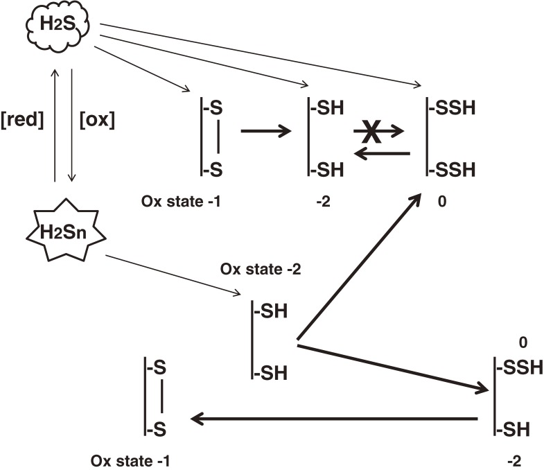
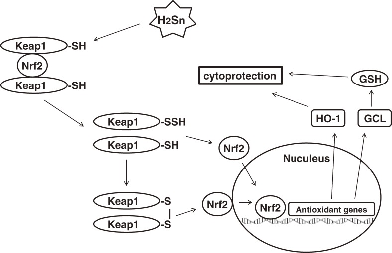
Hydrogen sulfide (H2S) is a familiar toxic gas that smells of rotten eggs. After the identification of endogenous H2S in the mammalian brain two decades ago, studies of this molecule uncovered physiological roles in processes such as neuromodulation, vascular tone regulation, cytoprotection against oxidative stress, angiogenesis, anti-inflammation, and oxygen sensing. Enzymes that produce H2S, such as cystathionine β-synthase, cystathionine γ-lyase, and 3-mercaptopyruvate sulfurtransferase have been studied intensively and well characterized. Polysulfides, which have a higher number of inner sulfur atoms than that in H2S, were recently identified as potential signaling molecules that can activate ion channels, transcription factors, and tumor suppressors with greater potency than that of H2S. This article focuses on our contribution to the discovery of these molecules and their metabolic pathways and mechanisms of action.


Figures

Figure 1.
Figure 1.
H2S facilitates the induction of hippocampal long-term potentiation (LTP) by enhancing the activity of NMDA receptors. A. A weak-tetanic stimulation, which alone does not induce LTP, induces LTP in the presence of H2S. B and C. H2S enhances the activity of NMDA receptors but not that of AMPA receptors, another type of ionotropic glutamate receptors. D. A lower concentration of NaHS, a sodium salt of H2S, further facilitated the induction of LTP even after DTT treatment. (Figures in 19 were modified).
Figure 2.
Figure 2.
The expression of CBS in the brain and the production of H2S. A. CBS is expressed in the brain. B. The production of H2S is suppressed by hydroxylamine (NH2OH) and aminooxyacetate (AOA), inhibitors of CBS, but not by propargylglycine (PGly), an inhibitor of CSE. S-adenosyl methionine (AdoMet) enhances the production of H2S (Figures in 19 were modified).
Figure 3.
Figure 3.
A synergistic effect of H2S with NO on vascular smooth muscle relaxation. The vascular relaxation effect of sodium nitroprusside (A) and morpholinosydnonimine (B) is greatly enhanced in the presence of H2S (Figures in 23 were modified). C. H2S, which is produced by 3MST together with CAT in endothelium and CSE in smooth muscle, relaxes smooth muscle and hyperpolarizes the membrane potential by activating potassium channels.
Figure 4.
Figure 4.
H2S passes through the membrane and HS through HS channels. H2S dissociates to HS and H+ with a trace amount of S2− under physiological conditions. At pH 7.4 and 37 ℃, approximately 20% H2S exists as a gas, and remaining 80% as HS. In mammalian cells, H2S passes through the plasma membrane and then dissociates under the extracellular environment where pH is slightly higher than inside the cell. The anion exchange protein AE1 transports HS in exchange for Cl. In bacteria, HS is released through HS channels to the extracellular environment, while H2S enters into cells through the plasma membrane similar to that seen for mammalian cells.
Figure 5.
Figure 5.
Two forms of intracellular sulfur that can release H2S. The iron-sulfur cluster, which localizes to the active center of enzymes in the respiratory chain and releases H2S under acidic conditions, forms the major acid-labile sulfur in cells. Bound sulfane sulfur, which consists of polysulfide and persulfide bound to proteins, releases H2S under reducing conditions and may form the intracellular storage for H2S.
Figure 6.
Figure 6.
H2S-producing pathways. CBS and CSE are localized to cytosol and produce H2S from l-cysteine alone or l-cysteine along with l-homocysteine. 3MST and CAT are localized to both cytosol and mitochondria. l-cysteine and α–ketoglutarate are metabolized by CAT to 3-MP, which is a substrate for 3MST to produce H2S. Thioredoxin interacts with 3MST to produce H2S (see also Fig. 7). d-Cysteine is metabolized in peroxisomes by DAO to 3MP, which is transported into mitochondria via a specific form of the vesicular transport. In mitochondria, 3MP is metabolized by 3MST to H2S. Peroxisome and mitochondria are in close vicinity or have a physical contact.
Figure 7.
Figure 7.
Transcription factor SP-1 up-regulates the CSE gene. H2S produced by CSE, whose transcription is increased by TGF-α mediated through SP1 binding activation, sulfurates (sulfhydrates) NF-κB to make it translocate into the nucleus and up-regulate antiapoptotic genes. Increased production of CSE by the activation of SP1 also plays an important role in the vascular smooth muscle differentiation.
Figure 8.
Figure 8.
3MST produces H2S with thioredoxin as an acceptor of sulfane sulfur. 3MST receives sulfur from 3MP to produce 3MST persulfide, which is transferred to one of the thiols in thioredoxin to generate thioredoxin persulfide. The remaining thiol reacts with persulfide to release H2S. Oxidized thioredoxin is reduced back to its reduced form by thioredoxin reductase.
Figure 9.
Figure 9.
Cytoprotective effect of H2S. H2S protects embryonic neurons from oxidative stress induced by high concentrations of glutamate. H2S enhances the activity of the cystine/glutamate antiporter and the cysteine transporter to increase the intracellular concentrations of cysteine. H2S also enhances the activity of glutamate cysteine lygase (GCL) to produce γ–glutamylcysteine to which glycine is added by glutathione synthetase (GC) to generate glutathione (GSH). H2S produced via the 3-mercaptopyruvate sulfurtransferase (3MST)/(CAT) pathway scavenges reactive oxygen species (ROS), which are abundantly generated in mitochondria. The intracellular concentrations of GSH are 1–10 mM, while those of H2S are only 10 nM to 3 µM. H2S efficiently suppresses oxidative stress by increasing the production of GSH rather than by scavenging ROS.
Figure 10.
Figure 10.
Existence of H2Sn in the brain and the induction of Ca2+ influx in astrocytes. H2S3 induces Ca2+ influx in astrocdytes in a dose-dependent manner with EC50 = 91 nM (A and B). Because H2Sn are mixture of molecules with different number of sulfur atoms in equilibrium as shown in the Eq. [14], even the standard Na2S3 and Na2S4 exert several peaks at the same retention times. Brain samples contain H2Sn (Figures in 120 were modified).
Figure 11.
Figure 11.
H2Sn activates TRPA1 channels. TRPA1 channels are activated by H2Sn by sulfurating either one or both of the two cysteine residues at their amino terminus, which then form cysteine disulfide bonds. The conformational changes in TRPA1 channels induce Ca2+ influx.
Figure 12.
Figure 12.
H2S together with H2Sn facilitates the induction of LTP. H2S enhances the activity of NMDA receptors by reducing a cysteine disulfide bond at the hinge of the ligand-binding domain of the receptors. H2Sn activates TRPA1 channels in astrocytes to induce Ca2+ influx, which facilitates the release of the gliotransmitter, d-serine, to the synaptic cleft. d-serine enhances the activity of NMDA receptors. By these effects of H2S and H2Sn LTP is effectively induced.
Figure 13.
Figure 13.
H2S reduces cysteine disulfide bond while H2Sn sulfurates cysteine residues. The atoms with the same oxidation state are not able to transfer electrons and take part in a redox reaction. H2S reduces sulfur in the cysteine disulfide bond (oxidation state −1) and that in persulfide (oxidation state 0) to cysteine residues (oxidation state −2), while H2Sn (oxidation state 0) sulfurates cysteine residues. The resulting persulfide reacts with thiols (oxidation state −2) to produce cysteine disulfide bonds.
Figure 14.
Figure 14.
H2Sn facilitates the nuclear translocation of Nrf2 to up-regulate antioxidant genes. H2Sn sulfurates Keap1 to release Nrf2, which translocates to the nucleus and up-regulates the transcription of antioxidant genes such as heme oxygenase 1 and glutamate cysteine ligase to increase the production of glutathione. By these effects on Keap1/Nrf2 complex, H2Sn exerts its cytoprotective activity.

Similar articles

Cited by

References

    1. Furchgott R.F., Zawadzki J.V. (1980) The obligatory role of endothelial cells in the relaxation of arterial smooth muscle by acetylcholine. Nature 288, 373–376. - PubMed
    1. Furchgott, R.F. (1988) Studies on relaxation of rabbit aorta by sodium nitrate: basis for the proposal that the acid-activatable component of the inhibitory factor from retractor penis is inorganic nitrate and the endothelium-derived relaxing factor is nitric oxide. In Mechanisms of Vasodilatation (ed. Vanhoutte, P.M.). Raven, New York, pp. 401–414.
    1. Ignarro, L.J., Byrns, R.E. and Wood, K.S. (1988) Biochemical and pharmacological properties of endothelium-derived relaxing factor and its similarity to nitric oxide radical. In Mechanisms of Vasodilatation (ed. Vanhoutte, P.M.). Raven, New York, pp. 427–435.
    1. Garthwaite J., Charles S.L., Chess-Williams R. (1988) Endothelium-derived relaxing factor release on activation of NMDA receptors suggests role as intercellular messenger in the brain. Nature 336, 385–388. - PubMed
    1. O’Dell T.J., Hawkins R.D., Kandel E.R., Arancio O. (1991) Tests of the roles of two diffusible substances in long-term potentiation: evidence for nitric oxide as a possible early retrograde messenger. Proc. Natl. Acad. Sci. U.S.A. 88, 11285–11289. - PMC - PubMed

Publication types