. 2015 May;57(4):506-17.
doi: 10.1002/dev.21305. Epub 2015 Apr 11.

A valuable and promising method for recording brain activity in behaving newborn rodents

Affiliations

A valuable and promising method for recording brain activity in behaving newborn rodents

Mark S Blumberg et al. Dev Psychobiol. 2015 May.

Abstract

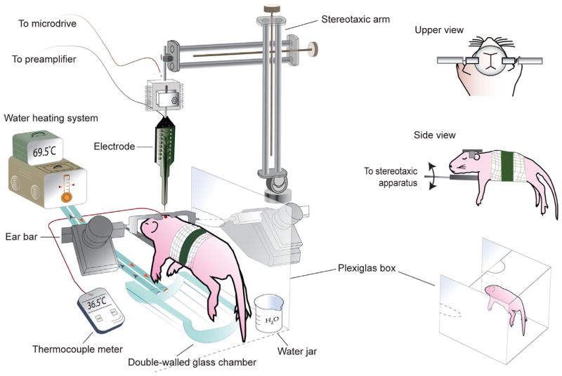
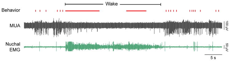
Neurophysiological recording of brain activity has been critically important to the field of neuroscience, but has contributed little to the field of developmental psychobiology. The reasons for this can be traced largely to methodological difficulties associated with recording neural activity in behaving newborn rats and mice. Over the last decade, however, the evolution of methods for recording from head-fixed newborns has heralded a new era in developmental neurophysiology. Here, we review these recent developments and provide a step-by-step primer for those interested in applying the head-fix method to their own research questions. Until now, this method has been used primarily to investigate spontaneous brain activity across sleep and wakefulness, the contributions of the sensory periphery to brain activity, or intrinsic network activity. Now, with some ingenuity, the uses of the head-fix method can be expanded to other domains to benefit our understanding of brain-behavior relations under normal and pathophysiological conditions across early development.

Keywords: EEG; imaging; infant; local field potential; mouse; multiunit activity; neurophysiology; pathophysiology; plasticity; rat; sleep; spontaneous activity; wake.


Figures

FIGURE 1
FIGURE 1
Constructing a head-fix device for infant rats and mice. Left: Materials needed to construct the device, including a large metal washer (A), metal strips (B), bolts (C), small metal washers (D), and nuts (E). The part numbers, dimensions, and prices of each part are provided in Table 2. The dimensions indicated for the large washer and metal strips are appropriate for use with 6- to 8-day-old rats. Right: Step-by-step method for constructing the device. (1) A hole is drilled at one end of each metal strip. The location of the hole determines the elevation of the pup’s head. (2) The end the strip containing the hole is bent 90° using flat-nose pliers. These operations must be performed identically on the two metal strips. (3) A bolt is secured with a washer (D) and nut (E) in each metal strip. The bolt should be further secured using cyanoacrylate adhesive. (4) The two metal strips are glued to the top of the washer. When performing this step, the bolts attached to each metal strip should be accurately aligned.
FIGURE 2
FIGURE 2
Recording set-up for head-fixed infant rats and mice. The head-fixed pup is secured on an elevated platform by applying tape around the gauze-wrapped torso and the platform. Ear bars are attached to the head-fix device and are secured to the stereotaxic apparatus. The double-walled glass chamber, through which temperature-controlled water is circulated, is positioned under the pup. To monitor temperature, a thermocouple is inserted through pre-drilled hole in the skull distant from the recording site. A Plexiglas box surrounds the pup on three sides to help maintain air temperature. A water-filled jar near the pup helps to maintain adequate humidity. An electrode, attached to a headstage on a stereotaxic arm, can be lowered into the brain via a pre-drilled hole using a microdrive.
FIGURE 3
FIGURE 3
Representative behavioral and electrophysiological records in a head-fixed week-old rat. The figure depicts nuchal EMG (green), multiunit activity (MUA, black), and behavior (red; vertical ticks: myoclonic twitches; horizontal lines: wake-related movements). Note the increased nuchal muscle tone during wake and the preceding and following periods of muscle atonia; the EMG spikes that occur against a background of atonia are twitches. (Adapted from Tiriac et al. (2014). Self-generated movements with “unexpected” sensory consequences. Current Biology, 24, 2136–2141, with permission.)

Similar articles

Cited by

References

    1. Ackman JB, Burbridge TJ, Crair MC. Retinal waves coordinate patterned activity throughout the developing visual system. Nature. 2012;490:219–225. - PMC - PubMed
    1. Alberts JR. Huddling by rat pups: Group behavioral mechanisms of temperature regulation and energy conservation. Journal of Comparative and Physiological Psychology. 1978;92:231–245. - PubMed
    1. Alberts JR, Blass E, Cramer C. Ecology and experience, sources of means and meaning of developmental change. In: Blass EM, editor. Handbook of behavioral neurobiology. Vol. 8. New York: Handbook of Behavioral Neurobiology; 1988. pp. 1–39.
    1. An S, Kilb W, Luhmann HJ. Sensory-evoked and spontaneous gamma and spindle bursts in neonatal rat motor cortex. Journal of Neuroscience. 2014;34:10870–10883. - PMC - PubMed
    1. An S, Yang JW, Sun H, Kilb W, Luhmann HJ. Long-term potentiation in the neonatal rat barrel cortex in vivo. Journal of Neuroscience. 2012;32:9511–9516. - PMC - PubMed

Publication types

LinkOut - more resources