

Evaluation of neurogenic potential of human umbilical cord mesenchymal cells; a time- and concentration-dependent manner
- PMID: 25864812
- PMCID: PMC4412918
- DOI: 10.6091/ibj.1452.2015
Evaluation of neurogenic potential of human umbilical cord mesenchymal cells; a time- and concentration-dependent manner
Abstract
Background: Retinoic acid as one of the most important regulators for cell differentiation was examined in this study for differentiation of human umbilical mesenchymal cells (hUCM).
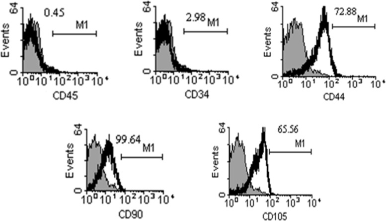
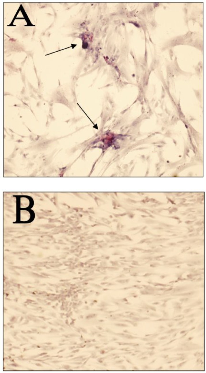
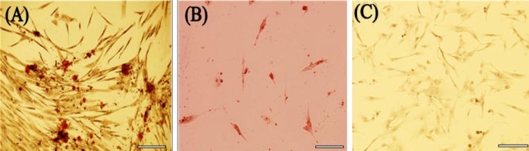
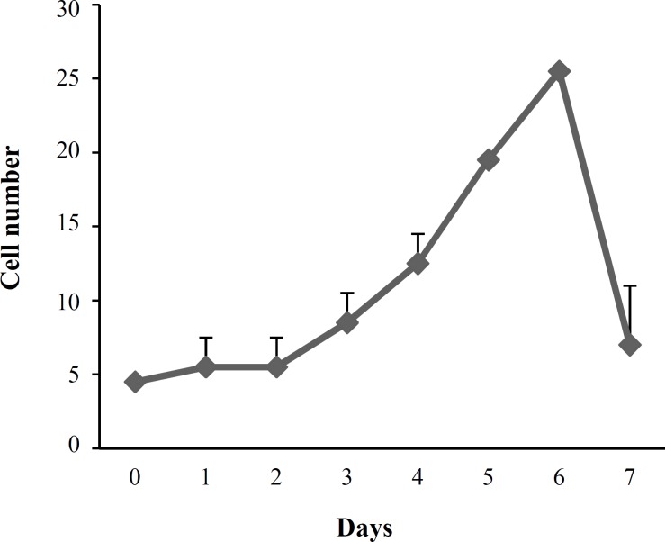
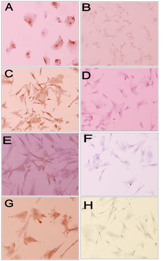
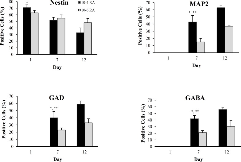
Methods: After isolation, hUCM were evaluated for mesenchymal stem cell properties by flow cytometry and alkaline phosphatase assay. Also, doubling time of the cells and their differentiation potential into adipogenic and osteogenic cells were tested. hUCM were then cultured with different concentrations of retinoic acid, and on days 1, 7, and 12, the percentage of differentiated cells was determined by immunostaining for nestin, anti-microtubule associated protein 2 (MAP₂), glutamic acid decarboxylase (GAD), and gamma-aminobutyric acid (GABA) markers.
Results: The isolated cells were negative for the hematopoietic markers and positive for the mesenchymal markers. They showed the population doubling time 60 ± 3 hours and differentiated into osteogenic and adipogenic cells. A descending trend in nestin and an ascending trend in MAP₂, GAD, and GABA expression were observed from the first day until the last day between different concentrations of retinoic acid.
Conclusion: hUCM cells may have the potential to differentiate into neural cells in the presence of different incubation period and concentration of retinoic acid.
Keywords: Cell differentiation; Neural stem cells; Retinoic acid.
Figures
Similar articles
-
The order of green and red LEDs irradiation affects the neural differentiation of human umbilical cord matrix-derived mesenchymal cells.Sci Rep. 2024 Sep 10;14(1):21107. doi: 10.1038/s41598-024-72099-3. Sci Rep. 2024. PMID: 39256554 Free PMC article.
-
Effects of light emitting diode irradiation on neural differentiation of human umbilical cord-derived mesenchymal cells.Sci Rep. 2017 Aug 30;7(1):9976. doi: 10.1038/s41598-017-10655-w. Sci Rep. 2017. PMID: 28855704 Free PMC article.
-
Calcium-sensing receptor-mediated osteogenic and early-stage neurogenic differentiation in umbilical cord matrix mesenchymal stem cells from a large animal model.PLoS One. 2014 Nov 7;9(11):e111533. doi: 10.1371/journal.pone.0111533. eCollection 2014. PLoS One. 2014. PMID: 25379789 Free PMC article.
-
[Biological characteristics of human umbilical cord-derived mesenchymal stem cells and their differentiation into neurocyte-like cells].Zhonghua Er Ke Za Zhi. 2006 Jul;44(7):513-7. Zhonghua Er Ke Za Zhi. 2006. PMID: 17044977 Chinese.
-
PPARγ and Wnt Signaling in Adipogenic and Osteogenic Differentiation of Mesenchymal Stem Cells.Curr Stem Cell Res Ther. 2016;11(3):216-25. doi: 10.2174/1574888x10666150519093429. Curr Stem Cell Res Ther. 2016. PMID: 25986621 Review.
Cited by
-
An Overview on Mesenchymal Stem Cells Derived from Extraembryonic Tissues: Supplement Sources and Isolation Methods.Stem Cells Cloning. 2020 Jul 7;13:57-65. doi: 10.2147/SCCAA.S248519. eCollection 2020. Stem Cells Cloning. 2020. PMID: 32753904 Free PMC article. Review.
-
The order of green and red LEDs irradiation affects the neural differentiation of human umbilical cord matrix-derived mesenchymal cells.Sci Rep. 2024 Sep 10;14(1):21107. doi: 10.1038/s41598-024-72099-3. Sci Rep. 2024. PMID: 39256554 Free PMC article.
-
Effects of light emitting diode irradiation on neural differentiation of human umbilical cord-derived mesenchymal cells.Sci Rep. 2017 Aug 30;7(1):9976. doi: 10.1038/s41598-017-10655-w. Sci Rep. 2017. PMID: 28855704 Free PMC article.
-
Exploring the impact of acetylsalicylic acid and conditioned medium obtained from mesenchymal cells, individually and in combination, on cognitive function, histological changes, and oxidant-antioxidant balance in male rats with hippocampal injury.Brain Behav. 2024 Sep;14(9):e70010. doi: 10.1002/brb3.70010. Brain Behav. 2024. PMID: 39262160 Free PMC article.
References
-
- Lu Z, Zhao h, Xu J, Zhang Z, Zhang X, Zhang Y, et al. Human umbilical cord mesenchymal stem cells in the treatment of secondary progressive multiple sclerosis. J Stem Cell Res Ther. 2013;S6
-
- Peng J, Wang Y, Zhang L, Zhao B, Zhao Z, Chen J, et al. Human umbilical cord Wharton's jelly-derived mesenchymal stem cells differentiate into a Schwann-cell phenotype and promote neurite outgrowth in vitro. Brain Res Bull. 2011 Feb;84(3):235–43. - PubMed
-
- Yıldırım S, Balcı D, Akpınar P, Can A. Differentiation potentials of two stroma-resident tissue-specific stem cells. Niche. 2012;1:1–7.
-
- Zhang L, Tan X, Dong C, Zou L, Zhao H, Zhang X, et al. In vitro differentiation of human umbilical cord mesenchymal stem cells (hUCMSCs), derived from Wharton's jelly, into choline acetyltransferase (ChAT)-positive cells. Int J Dev Neurosci. 2012;30(6):471–7. - PubMed
-
- Ross SA, McCaffery PJ, Drager UC, De Luca LM. Retinoids in embryonal development. Physiol Rev. 2000 Jul;80(3):1021–54. - PubMed
Publication types
MeSH terms
Substances
LinkOut - more resources
Full Text Sources