. 2015 Apr 10:4:26334.
doi: 10.3402/jev.v4.26334. eCollection 2015.

Exosomal sorting of the viral oncoprotein LMP1 is restrained by TRAF2 association at signalling endosomes

Affiliations

Exosomal sorting of the viral oncoprotein LMP1 is restrained by TRAF2 association at signalling endosomes

Frederik J Verweij et al. J Extracell Vesicles. .

Abstract

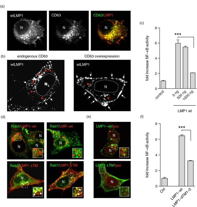
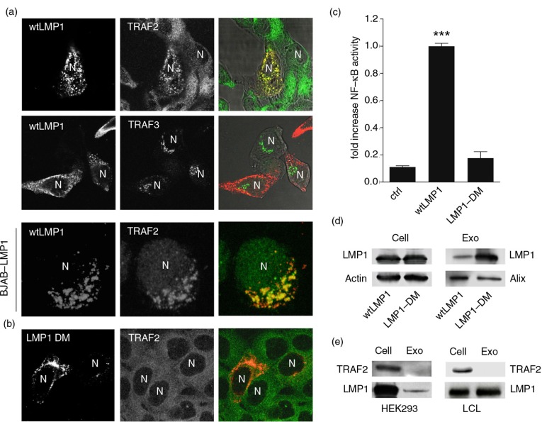
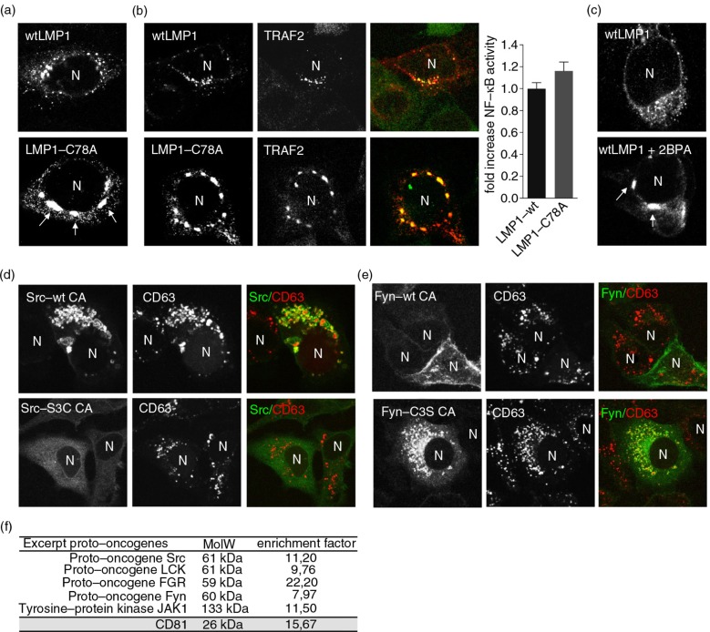
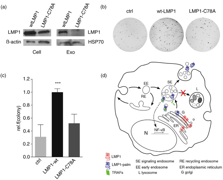
The Epstein-Barr virus (EBV)-encoded oncoprotein latent membrane protein 1 (LMP1) constitutively activates nuclear factor κB (NFκB) from intracellular membranes to promote cell growth and survival. LMP1 associates with CD63 in intracellular membranes and is released via exosomes. Whether tumour necrosis factor (TNF) receptor-associated factors (TRAFs) mediate LMP1 NFκB signalling from endosomes and modulate exosomal sorting is unknown. In this article, we show that LMP1-TRAF2 signalling complexes accumulate at endosomes in a palmitoylation-dependent manner, thereby driving LMP1-dependent oncogenicity. Palmitoylation is a reversible post-translational modification and is considered to function as a membrane anchor for proteins. Mutagenesis studies showed that LMP1-TRAF2 trafficking to endosomes is dependent on one single cysteine residue (C78), a known palmitoylation site of LMP1. Notably, growth assays in soft agar revealed that oncogenic properties of the palmitoylation-deficient LMP1 mutant C78A were diminished compared to wild-type LMP1. Since LMP1 recruitment of TRAF2 and downstream NFκB signalling were not affected by a disturbance in palmitoylation, the specific localization of LMP1 at endosomal membranes appears crucial for its transforming potential. The importance of palmitoylation for trafficking to and signalling from endosomal membranes was not restricted to LMP1, as similar observations were made for the cellular oncoproteins Src and Fyn. Despite abundant LMP1-TRAF2 association at endosomal membranes TRAF2 could not be detected in exosomes by Western blotting or proteomics. Interestingly, point mutations that prevented TRAF binding strongly promoted the sorting and release of LMP1 via exosomes. These observations reveal that LMP1-TRAF2 complexes at endosomes support oncogenic NFκB activation and suggest that LMP1 dissociates from the activated signalling complexes upon sorting into intraluminal vesicles. We propose that "signalling endosomes" in EBV-infected tumour cells can fuse with the plasma membrane, explaining LMP1 release via exosomes.

Keywords: LMP1; endosomes; exosomes; oncogenic signalling; palmitoylation.


Figures

Fig. 1
Fig. 1
LMP1 accumulates at and signals from CD63+ endosomes. (a) Immunofluorescent labelling of wtLMP1 (red) and CD63 (green) in HEK293 cells. (b) Immunofluorescent labelling of wtLMP1 in HEK293 cells with endogenous CD63 levels or overexpression of CD63. White and red arrowheads indicate LMP1 localized at the plasma membrane or endosomal membranes, respectively, and N indicates nucleus. (c) Reporter assay for effect of CD63 on LMP1-wt NFκB activity. Cell lysates of HEK293 cells transfected for 24 hours with wtLMP1 (LMP1-WT) and increasing amounts of CD63 plasmid or empty vector (control), together with an NFκB–reporter construct. Error bars represent s.d.; shown is one representative experiment; n=3. (d) Immunofluorescent labelling of LMP1-wt or LMP1ΔTM1-2 (LMP1 ΔTM) (both in red) in HEK293 cells co-transfected with Rab5- or Rab7-GFP (both in green). N indicates nucleus. (e) Immunofluorescent labelling of Lysotracker (red) in LMP1-wt or LMP1 ΔTM (green) transfected HEK293 cells. N indicates nucleus. (f) Reporter assay for LMP1-wt or LMP1-ΔTM1-2 NFκB activity. Cell lysates of HEK293 cells transfected with wtLMP1 (LMP1-WT), LMP1-ΔTM1-2 (LMP1-ΔTM1-2), or empty vector (control), together with an NFκB–reporter construct. Error bars represent s.d.; shown is one representative experiment; n=3.
Fig. 2
Fig. 2
LMP1–TRAF2 association at endosomal membranes controls signalling and sorting. (a) Immunofluorescent labelling of endogenous TRAF2 and TRAF3 (all in green) in wtLMP1 (red) transfected HEK293 cells. Lower panel: Immunofluorescent labelling on EBV-negative BJAB cells carrying a tetracycline-inducible (TET-Off) LMP1 expression construct, induced for 24 hours (LMP1 in red) and labelled for endogenous TRAF2 (green). N indicates nucleus. (b) Immunofluorescent labelling of endogenous TRAF2 (green) in HEK293 cells transfected with LMP1 constructs with point mutations in (LMP1 DM) the suspected TRAF-binding sites (red). N indicates nucleus. (c) Reporter assay for LMP1-wt or LMP1-DM NFκB activity. Cell lysates of HEK293 cells transfected for 24 hours with wtLMP1, LMP1-DM, or empty vector (control), together with an NFκB–reporter construct. Error bars represent s.d.; shown is one representative experiment; n=3. (d) Western blotting analysis on TRAF2 and LMP1 protein levels in EBV-infected LCL cell and exosome lysates (left) and in wtLMP1-transfected HEK293 cell and exosome lysates (right). (e) Western blotting analysis on LMP1 protein levels in wtLMP1– or LMP1-DM-transfected HEK293 cell and exosome lysates.
Fig. 3
Fig. 3
Palmitoylation controls subcellular trafficking of LMP1 but not TRAF2 association. (a) Immunofluorescent co-labelling of transfected wtLMP1 or LMP1-C78A (red) in HEK293 cells. N indicates nucleus. (b) Immunofluorescent labelling of endogenous TRAF2 (green) in HEK293 cells transfected with wtLMP1 or LMP1-C78A (red). To the right, reporter assay for LMP1-wt or LMP1-C78A NFκB activity. Cell lysates of HEK293 cells transfected for 24 hours with wtLMP1 or LMP1-C78A, together with an NFκB–reporter construct. Error bars represent s.e.m.; n>3. N indicates nucleus. (c) Immunofluorescent labelling of transfected wtLMP1 in 2BPA and control treated HEK293 cells. N indicates nucleus. (d, e) Immunofluorescent labelling of constitutive active Src-wt CA, Src-S3C CA, Fyn-wt CA, or Fyn-C3S CA (all in green) transfected HeLa-CIITA cells, co-labelled for CD63 (red). N indicates nucleus. (f) Table showing the enrichment of proto-oncogenes in exosomes versus cell lysates from 6 different B-cell lines referenced to the total number of peptides identified in each analysis.
Fig. 4
Fig. 4
Palmitoylation controls LMP1 transformation capacity. (a) Western blotting analysis on LMP1 protein levels in wtLMP1 or LMP1-C78A transfected HEK293 cell and exosome lysates; β-actin and HSP70 as loading controls. (b, c) Soft-agar assay for anchorage-independent growth of HEK293 cells transfected with wtLMP1, LMP1-C78A, or GFP control (ctrl) construct. Error bars represent s.e.m.; n>3. (d) Hypothetical model showing that newly synthesized LMP1 (blue) assembles with CD63 in the ER; traffics to the Golgi (G), where it is palmitoylated (red); and buds off in CD63-positive transport vesicles that form or assemble at limiting membranes of signalling endosomes (SEs). Whereas palmitoylation targets LMP1 to SE membranes, non-palmitoylated LMP1 is retained in the ER–Golgi region. Wild-type LMP1 recruits TRAF2, and the activated signalling complexes accumulate at endosomal membranes activating NFκB. Upon LMP1 sorting into the intra-luminal vesicles, LMP1 is then secreted via exosomes while TRAF2 dissociates and is presumably left behind in the cytosol.

References

    1. Scita G, Di Fiore PP. The endocytic matrix. Nature. 2010;463:464–73. - PubMed
    1. Joffre C, Barrow R, Menard L, Calleja V, Hart IR, Kermorgant S. A direct role for Met endocytosis in tumorigenesis. Nat Cell Biol. 2011;13:827–37. - PubMed
    1. Peinado H, Aleckovic M, Lavotshkin S, Matei I, Costa-Silva B, Moreno-Bueno G, et al. Melanoma exosomes educate bone marrow progenitor cells toward a pro-metastatic phenotype through MET. Nat Med. 2012;18:883–91. - PMC - PubMed
    1. "/>Al-Nedawi K, Meehan B, Micallef J, Lhotak V, May L, Guha A, et al. Intercellular transfer of the oncogenic receptor EGFRvIII by microvesicles derived from tumour cells. Nat Cell Biol. 2008;10:619–24. - PubMed
    1. Sandilands E, Frame MC. Endosomal trafficking of Src tyrosine kinase. Trends Cell Biol. 2008;18:322–9. - PubMed