

Entosis allows timely elimination of the luminal epithelial barrier for embryo implantation
- PMID: 25865893
- PMCID: PMC5089169
- DOI: 10.1016/j.celrep.2015.03.035
Entosis allows timely elimination of the luminal epithelial barrier for embryo implantation
Abstract
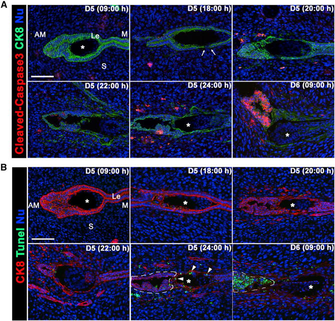
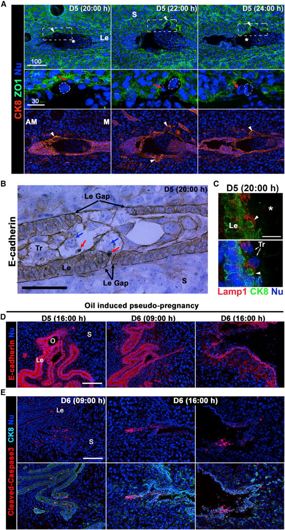
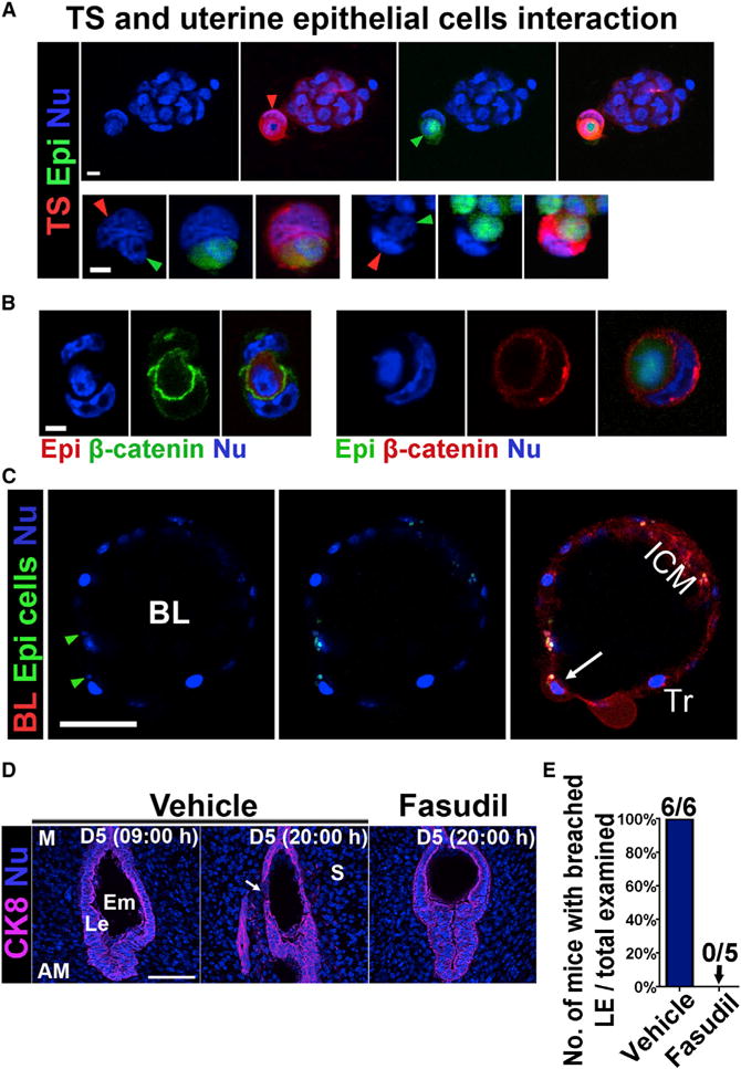
During implantation, uterine luminal epithelial (LE) cells first interact with the blastocyst trophectoderm. Within 30 hr after the initiation of attachment, LE cells surrounding the blastocyst in the implantation chamber (crypt) disappear, allowing trophoblast cells to make direct physical contact with the underneath stroma for successful implantation. The mechanism for the extraction of LE cells was thought to be mediated by apoptosis. Here, we show that LE cells in direct contact with the blastocyst are endocytosed by trophoblast cells by adopting the nonapoptotic cell-in-cell invasion process (entosis) in the absence of caspase 3 activation. Our in vivo observations were reinforced by the results of co-culture experiments with primary uterine epithelial cells with trophoblast stem cells or blastocysts showing internalization of epithelial cells by trophoblasts. We have identified entosis as a mechanism to remove LE cells by trophoblast cells in implantation, conferring a role for entosis in an important physiological process.
Copyright © 2015 The Authors. Published by Elsevier Inc. All rights reserved.
Figures

Similar articles
-
Differential expression of ezrin/radixin/moesin (ERM) and ERM-associated adhesion molecules in the blastocyst and uterus suggests their functions during implantation.Biol Reprod. 2004 Mar;70(3):729-36. doi: 10.1095/biolreprod.103.022764. Epub 2003 Nov 12. Biol Reprod. 2004. PMID: 14613898
-
Dual roles of progesterone in embryo implantation in mouse.Endocrine. 2003 Jul;21(2):123-32. doi: 10.1385/ENDO:21:2:123. Endocrine. 2003. PMID: 12897374
-
Zonula occludens-1 and E-cadherin are coordinately expressed in the mouse uterus with the initiation of implantation and decidualization.Dev Biol. 1999 Apr 15;208(2):488-501. doi: 10.1006/dbio.1999.9206. Dev Biol. 1999. PMID: 10191061
-
Trophoblast-uterine interactions in the first days of implantation: models for the study of implantation events in the human.Semin Reprod Med. 2000;18(3):255-63. doi: 10.1055/s-2000-12563. Semin Reprod Med. 2000. PMID: 11299964 Review.
-
Implantation mechanisms: insights from the sheep.Reproduction. 2004 Dec;128(6):657-68. doi: 10.1530/rep.1.00398. Reproduction. 2004. PMID: 15579583 Review.
Cited by
-
The investigation of the role of sirtuin-1 on embryo implantation in oxidative stress-induced mice.J Assist Reprod Genet. 2021 Sep;38(9):2349-2361. doi: 10.1007/s10815-021-02229-7. Epub 2021 May 15. J Assist Reprod Genet. 2021. PMID: 33993396 Free PMC article.
-
Molecular mechanisms underlying cell-in-cell formation: core machineries and beyond.J Mol Cell Biol. 2021 Aug 18;13(5):329-334. doi: 10.1093/jmcb/mjab015. J Mol Cell Biol. 2021. PMID: 33693765 Free PMC article. No abstract available.
-
Unconventional Ways to Live and Die: Cell Death and Survival in Development, Homeostasis, and Disease.Annu Rev Cell Dev Biol. 2018 Oct 6;34:311-332. doi: 10.1146/annurev-cellbio-100616-060748. Epub 2018 Aug 8. Annu Rev Cell Dev Biol. 2018. PMID: 30089222 Free PMC article. Review.
-
Establishing 3D Endometrial Organoids from the Mouse Uterus.J Vis Exp. 2023 Jan 6;(191):10.3791/64448. doi: 10.3791/64448. J Vis Exp. 2023. PMID: 36688555 Free PMC article.
-
Megakaryocyte emperipolesis: a new frontier in cell-in-cell interaction.Platelets. 2020 Aug 17;31(6):700-706. doi: 10.1080/09537104.2019.1693035. Epub 2019 Nov 21. Platelets. 2020. PMID: 31752579 Free PMC article. Review.
References
-
- Das SK, Wang XN, Paria BC, Damm D, Abraham JA, Klagsbrun M, Andrews GK, Dey SK. Heparin-binding EGF-like growth factor gene is induced in the mouse uterus temporally by the blastocyst solely at the site of its apposition: a possible ligand for interaction with blastocyst EGF-receptor in implantation. Development. 1994;120:1071–1083. - PubMed
Publication types
MeSH terms
Grants and funding
LinkOut - more resources
Full Text Sources
Other Literature Sources
Research Materials
