. 2015 Apr 16;58(2):255-68.
doi: 10.1016/j.molcel.2015.03.011. Epub 2015 Apr 9.

PTEN functions by recruitment to cytoplasmic vesicles

Affiliations

PTEN functions by recruitment to cytoplasmic vesicles

Adam Naguib et al. Mol Cell. .

Abstract

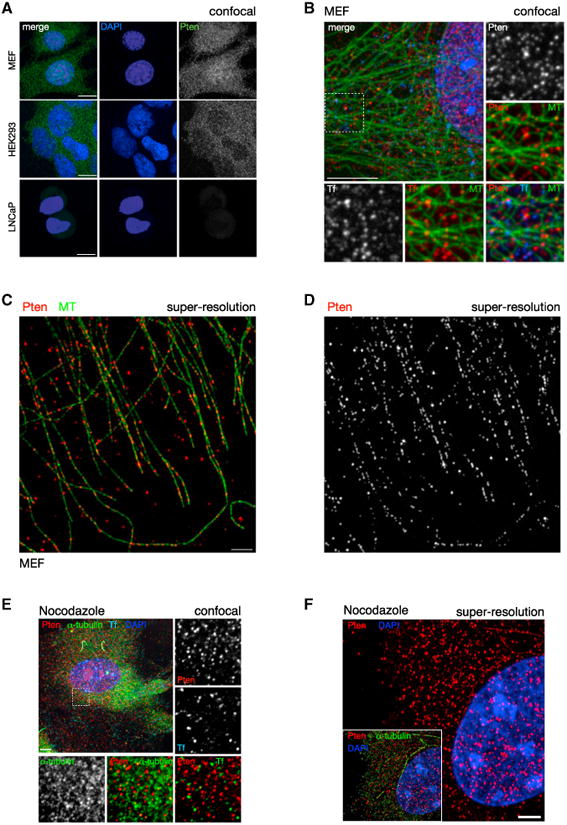
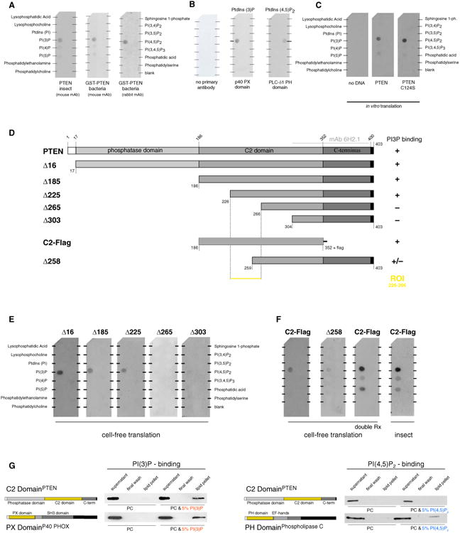
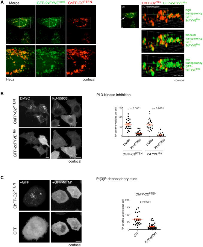
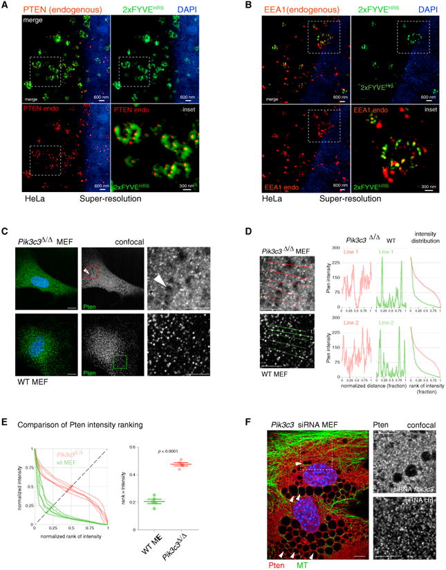
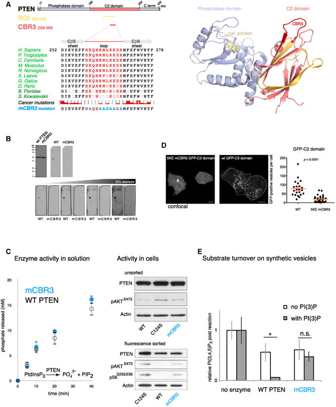
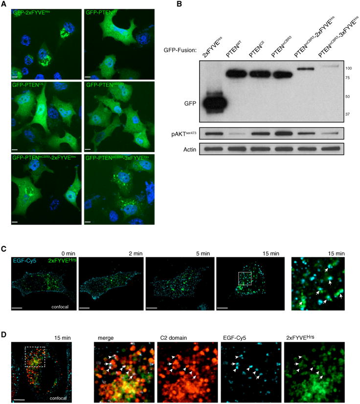
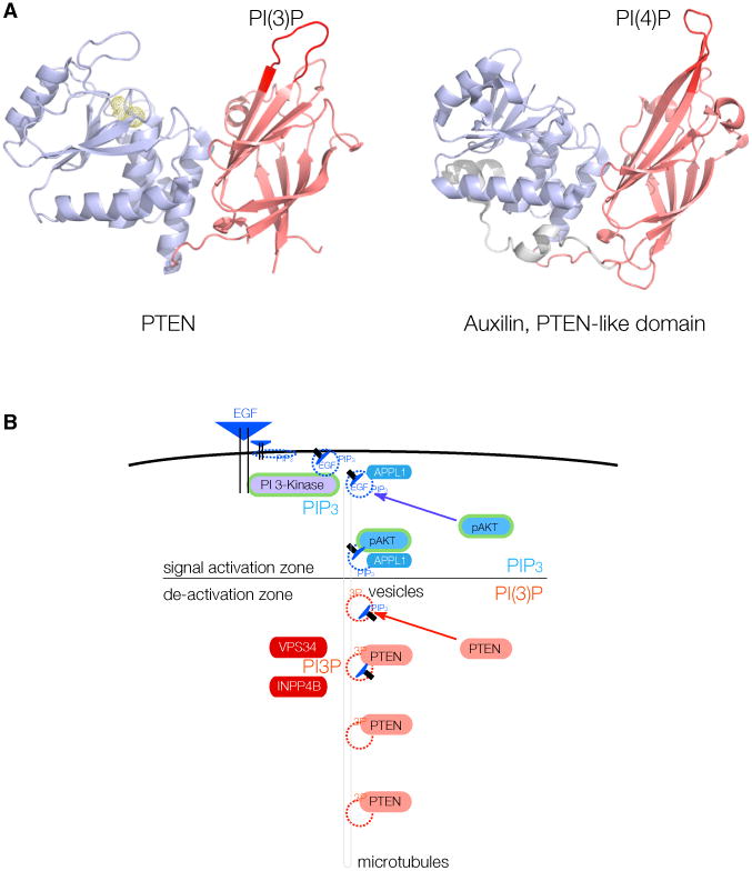
PTEN is proposed to function at the plasma membrane, where receptor tyrosine kinases are activated. However, the majority of PTEN is located throughout the cytoplasm. Here, we show that cytoplasmic PTEN is distributed along microtubules, tethered to vesicles via phosphatidylinositol 3-phosphate (PI(3)P), the signature lipid of endosomes. We demonstrate that the non-catalytic C2 domain of PTEN specifically binds PI(3)P through the CBR3 loop. Mutations render this loop incapable of PI(3)P binding and abrogate PTEN-mediated inhibition of PI 3-kinase/AKT signaling. This loss of function is rescued by fusion of the loop mutant PTEN to FYVE, the canonical PI(3)P binding domain, demonstrating the functional importance of targeting PTEN to endosomal membranes. Beyond revealing an upstream activation mechanism of PTEN, our data introduce the concept of PI 3-kinase signal activation on the vast plasma membrane that is contrasted by PTEN-mediated signal termination on the small, discrete surfaces of internalized vesicles.


Figures

Figure 1
Figure 1. PTEN Is Organized along MTs
(A) Confocal immunofluorescence (IF) microscopy reveals that PTEN distribution is punctate in the cytoplasm. Punctate distribution is conserved in both primary WT MEFs (top panels) and HEK293 cell lines (middle panels). PTEN null cells (human prostate cancer-derived LNCaP) display minimal background staining, confirming antibody specificity (bottom panels). Images show total projections of all Z sections (extended focus). Scale bars, 10 μm. (B) Cytoplasmic PTEN distribution is indistinguishable from that of labeled transferrin (Tf). Pten and Tf punctate stains show a similar close association with MTs. Images of WT MEFs, single section (Z = 1). Scale bar, 10 μm. (C and D) Super-resolution light microscopy (OMX) reveals that PTEN is organized along MTs. Scale bar, 2 μm. (E) Nocodazole treatment of WT MEFs abrogates MT assembly but does not dissolve the punctae of Pten or Transferrin, compared to alpha-tubulin. Z = 1. Scale bar, 10 μm. (F) Nocodazole treatment abrogates the linear arrangement of PTEN along MTs. Z = 1. Scale bar, 2 μm. See also Figure S1.
Figure 2
Figure 2. PTEN Directly Binds to the Endosomal Signature Lipid PI(3)P
(A) Recombinant human PTEN protein directly binds PI(3)P as shown by incubation of GST-PTEN and untagged PTEN (generated using the indicated protein expression systems) with immobilized lipids. Note that binding to PI(4)P and PI(5)P is weak and variable. mAb, monoclonal antibody. (B) Recombinant PX domain (from p40 PHOX) and PH domain (from phospholipase C-δ1) specifically recognize their respective lipid targets PI(3)P and PI(4,5)P2, while secondary antibody alone shows no staining. PtdIns, PI. (C) Cell-free translation of PTEN using bacterial extract reproduces PI(3)P binding of WT PTEN (middle) and the C124S catalytically inactive mutant (right). ph, phosphate. (D and E) Truncation maps (D) and overlay assays (E) performed with the indicated PTEN mutants identifies the ROI containing the minimal PI(3)P-binding element. (F) The C2 domain of PTEN in isolation (with a C-terminal flag epitope for antibody detection) is necessary and sufficient for binding to PI(3)P. Doubling of reaction volumes for cell-free translation yields comparable results to reactions performed with insect-cell-purified C2 domain of PTEN. (G) The C2 domain of PTEN binds synthetic liposomes containing 5% PI(3)P in a phosphatidylcholine (PC) membrane similar to the known PI(3)P-binding PX domain of the p40 PHOX protein. The C2 domain of PTEN does not interact with PI(4,5)P2 as shown using PC liposomes containing 5% PI(4,5)P2 and the known PI(4,5)P2-binding PH domain of phospholipase C as a positive control. See also Figure S2.
Figure 3
Figure 3. The C2 Domain of PTEN Is a Novel PI(3)P-Binding Domain
(A) Left panels: mCherry fluorescent protein (ChFP)-tagged PTEN C2 domain extensively co-localizes with the GFP-tagged FYVE domain of HRS protein, a prototypical PI(3)P probe in cells. Z = 1. Scale bar, 10 μm. Note that identical results are obtained with a GFP-tagged C2 domain (see Figures 5D and S2C). Right panels: 3D rendering of colocalization between PTEN C2 domain and 2×FYVEHRS. Arrow represents the angle of view. Three density settings are displayed for 2×FYVEHRS to allow complete visualization of the signal overlap with ChFP-C2PTEN domain. Unit length of the 3D floor is 9.5 μm. (B) The number of PTEN C2-domain-positive and 2×FYVEHRS-domain-positive vesicles is diminished in HeLa cells subsequent to treatment with the PI 3-K inhibitor KU-55933. Scale bars, 10 μm, extended focus. Graph, red bars represent the mean value, and the whiskers represent SEM. p < 0.0001, two-tailed Student's t test. (C) Overexpression of the PI(3)P phosphatase MTM1 (GFP-MTM1) suppresses C2PTEN-vesicle association. Scale bar, 10 μm, extended focus. Graph, red bars show mean with SEM, p < 0.0001, two-tailed Student's t test. See also Figure S3.
Figure 4
Figure 4. PTEN Localization Is Dictated by PI(3)P
(A) Super-resolution microscopy demonstrates colocalization of endogenous (endo) PTEN with overexpressed GFP-2×FYVEHRS, Z = 1. 2D, two-dimensional. (B) Similar results are obtained with endogenous EEA1 and overexpressed GFP-2×FYVEHRS, Z = 1. (C) Pten shows non-punctate cytoplasmic staining in Vps34 (Pik3c3 gene) knockout MEFs (see Experimental Procedures). Arrowheads indicate large vacuole formation, which confirms loss of Vps34 function. Z = 1. Scale bar, 10 μm. (D) Line intensity profiles of Pten in Pik3c3 null and WT MEFs from (C), Z =1. Scale bar, 10 μm. Right panels: absolute intensity is plotted versus normalized rank of absolute intensity. (E) Quantification of Pten intensity plots. Normalized intensity is plotted versus normalized rank of intensity on five lines shown. Comparison of a measure for rank: intensity distribution (see Experimental Procedures) shows significant difference between WT and knockout cells. Bars represent the mean value, and the whiskers represent SEM. p < 0.0001, two-tailed Student's t test. (F) siRNA against Vps34 (Pik3c3 gene) confirms loss of discrete Pten punctae in Vps34-knockdown MEFs. See Figures S3C and S3E for quantification. Arrowheads indicate vacuoles, confirming suppression of Vps34 function. Z = 1. Scale bars, 10 μm. Ctrl, control. See also Figure S3.
Figure 5
Figure 5. PI3P-Dependent Localization and Activation Is Mediated by the CBR3 Loop of PTEN
(A) Left: domain structure of PTEN with the identified ROI from Figure 2 (gold) and the CBR3 loop (red). Middle: conservation of the CBR3 loop (red) in the C2 domain among vertebrates and cancer-associated missense mutations. The top reported cancer mutation count (9) is found for the Trp-274 residue. Three cancer-associated missense mutations have been reported inside the CBR3 (Cerami et al., 2012). The last row shows mutations engineered to neutralize CBR3 loop function. Right: PTEN structure highlighting the CBR3 loop (red) in the ROI (gold). Note that the CBR3 is positioned to face the target membrane in concert with the catalytic pocket (Georgescu et al., 2000; Lee et al., 1999) (active-site cysteine is shown in yellow as ball-and-stick residue). (B) Mutation of the CBR3 loop (mCBR3 PTEN), as indicated in (A), abolishes PTEN-PI(3)P binding. (C) The mCBR3 PTEN mutant retains normal lipid phosphatase activity against soluble substrate (diC 8 PI(3,4,5)P3) (graph; error bars indicate SD, n = 2). Top: immunoblot analysis of overexpressed mCBR3 mutant GFP-PTEN reveals strongly impaired pathway antagonizing function compared to WT GFP-PTEN in PTEN-deficient PC3 cells. Bottom: immunoblot analysis revels that FACS-sorted mCBR3 mutant GFP-PTEN cells have higher AKT phosphorylation than cells transfected with WT GFP-PTEN. (D) GFP-tagged 5KE mutant C2 domain of PTEN is less efficiently recruited to PI(3)P-positive membranes than the WT(wt) GFP-tagged C2PTEN domain. Student's t test, two tailed. Z = 1. Scale bar, 10 μm. (E) Mass-spectrometry-based quantification of PI(3,4,5)P3 abundance shows that both WT and mCBR3 proteins are active on liposomes. More productive catalysis of WT enzyme is achieved by the addition of PI(3)P to liposome membranes, but, in contrast, no enhanced activity is observed with the mCBR3 enzyme. Error bars indicate SEM. *p < 0.05, Student's t test, two-tailed.
Figure 6
Figure 6. Targeting of PTEN to PI(3)P Is Necessary for Function in Cells
(A) Overexpression of multiple GFP-PTEN fusion proteins and their sub-cellular localization. Z = 1. Scale bar, 10 μm. (B) Overexpression of PTEN constructs followed by FACS and interrogation of AKT phosphorylation. Fusion of mCBR3 PTEN to FYVE domains restores the ability of PTEN to efficiently antagonize PI3-K/AKT-mediated signaling. (C) EGF-Cy5 internalization leads to colocalization with GFP-2×FYVEHRS after 15 min (arrows). Z = 1. Scale bar, 10 μm. (D) Colocalization of ChFP-C2PTEN domain with EGF-Cy5 (arrowheads) and of ChFP-C2PTEN domain, EGF-Cy5, and GFP-2×FYVEHRS (arrows) is seen at 15 min post-EGF-Cy5 internalization. Z = 1. Scale bar, 10 μm. See also Figure S4.
Figure 7
Figure 7. PTEN Vesicle Binding May Be an Evolutionarily Conserved Function in Endocytosis
(A) Left: phosphatase (blue) and C2 domain (red) of PTEN, with the essential CBR3 PI(3)P-binding loop highlighted (left, crimson). Right: structure of the PTEN-like region of Auxilin, with the essential loop for PI(4)P binding highlighted (right, crimson). (B) Model for spatio-temporal maturation of vesicle-associated PIP3-lipid signaling through APPL1-positive vesicles (signal activation zone)and subsequent PIP3 signal termination by PTEN after generation of PI(3)P (also labeled 3P) onto vesicle membranes by, e.g., VPS34 or INPP4B (de-activation zone).

Comment in

References

    1. Alimonti A, Carracedo A, Clohessy JG, Trotman LC, Nardella C, Egia A, Salmena L, Sampieri K, Haveman WJ, Brogi E, et al. Subtle variations in Pten dose determine cancer susceptibility. Nat Genet. 2010;42:454–458. - PMC - PubMed
    1. Backer JM. The regulation and function of Class III PI3Ks: novel roles for Vps34. Biochem J. 2008;410:1–17. - PubMed
    1. Berger AH, Knudson AG, Pandolfi PP. A continuum model for tumour suppression. Nature. 2011;476:163–169. - PMC - PubMed
    1. Bravo J, Karathanassis D, Pacold CM, Pacold ME, Ellson CD, Anderson KE, Butler PJ, Lavenir I, Perisic O, Hawkins PT, et al. The crystal structure of the PX domain from p40(phox) bound to phosphatidylinositol 3-phosphate. Mol Cell. 2001;8:829–839. - PubMed
    1. Butler MG, Dasouki MJ, Zhou XP, Talebizadeh Z, Brown M, Takahashi TN, Miles JH, Wang CH, Stratton R, Pilarski R, Eng C. Subset of individuals with autism spectrum disorders and extreme macrocephaly associated with germline PTEN tumour suppressor gene mutations. J Med Genet. 2005;42:318–321. - PMC - PubMed

Publication types

Substances