Review
. 2015;15(12):1138-52.
doi: 10.2174/1568026615666150413153400.

Applications of aptamers in targeted imaging: state of the art

Affiliations
Review

Applications of aptamers in targeted imaging: state of the art

Casey A Dougherty et al. Curr Top Med Chem. 2015.

Abstract

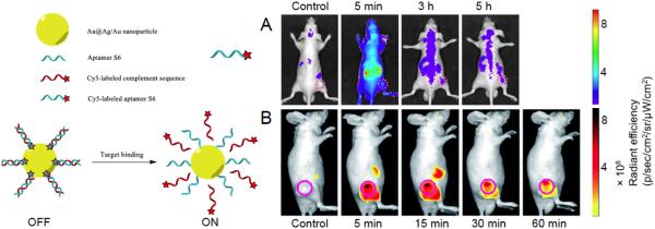
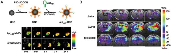
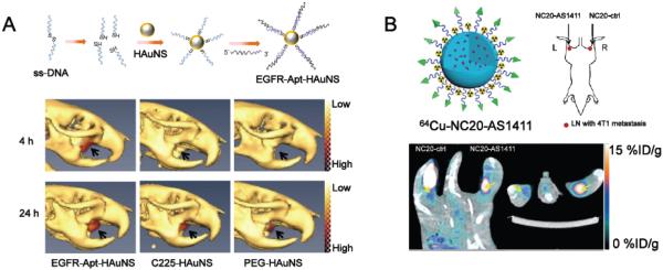
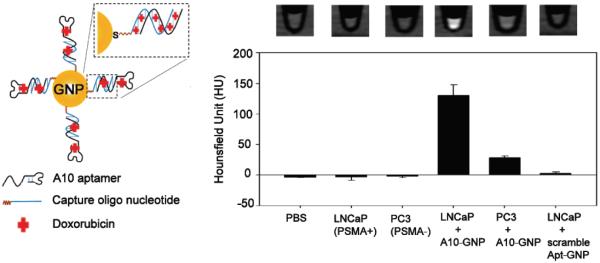
Aptamers are single-stranded oligonucleotides with high affinity and specificity to the target molecules or cells, thus they can serve as an important category of molecular targeting ligand. Since their discovery, aptamers have been rapidly translated into clinical practice. The strong target affinity/selectivity, cost-effectivity, chemical versatility and safety of aptamers are superior to traditional peptides- or proteins-based ligands which make them unique choices for molecular imaging. Therefore, aptamers are considered to be extremely useful to guide various imaging contrast agents to the target tissues or cells for optical, magnetic resonance, nuclear, computed tomography, ultrasound and multimodality imaging. This review aims to provide an overview of aptamers' advantages as targeting ligands and their application in targeted imaging. Further research in synthesis of new types of aptamers and their conjugation with new categories of contrast agents is required to develop clinically translatable aptamer-based imaging agents which will eventually result in improved patient care.


Figures

Fig. (1)
Fig. (1)
(A) Fluorescently labeled “always-on” S6 aptamer (for A549 targeting) for fluorescence imaging in A549 xenografts. Adapted with penni ssion from reference [30]. (B) The use of S6 aptamer-Au@Ag/Au nanoparticle based AAP for fluorescence imaging in A549 xenografts. Better tumor-to-background ratio was witnessed compared with directly labeled S6 aptamer. Adapted with petmission from reference [48].
Fig. (2)
Fig. (2)
(A) An integrin αvβ3 aptamer (Aptαvβ3) conjugated magnetic nanoparticle (MNP) for MRI of A341 xenografts (integrin αvβ3 +). Aptαvβ3-MNPs showed more persistent uptake in A341 tumors compared with cRGD-MNPs (cRGD is a well-adopted targeting ligand for integrin αvβ3). Pre: preinjection; Imm: immediately following the injections; MNC: magnetic nanocrystal. Adapted with permission from reference [65]. (B) The ΔR2* maps of C57B6 mice injected with 5ECdsAP1-SPIO for MRI detection of transcription factor AP1. Amphetamine (AMPH) is a strong stimulator of central nervous system which can result in the elevation of AP-1. SCH23390 is a dopaminergic receptor antagonist which can suppress the AP-1 elevation by AMPH. Saline was used as a control in this study. MRI was carried out 4 h post drug treatment and 7 h post intracerebroventricular injection of 5ECdsAP1-SPIO. Adapted with permission from reference [68].
Fig. (3)
Fig. (3)
(A) The structure and application of 111In-labeled, EGFR aptamer-conjugated HAuNS (EGFR-Apt-HAuNS) for SPECT/CT imaging in EGFR-positive OSC-19 xenografts. EGFR-Apt-HAuNS demonstrated faster penetration into OSC-19 tumors along with consistent accumulation when compared to EGFR-antibody (C225) conjugated HAuNS. Adapted with permission from reference [77]. (B) The use of 64Cu-labeled, AS1411-conjugated silica nanoparticles (NC20-AS1411) for PET imaging of 4T1 tumor-drained lymph nodes. 64Cu-labeled, unconjugated nanoparticles (NC20-ctrl) showed significantly lower uptake in these lymph nodes. Adapted with permission from reference [83].
Fig. (4)
Fig. (4)
PSMA-targeting A10 aptamer conjugated GNP for cellular CT imaging. Specific recognition of PSMA-positive LNCaP cells was witnessed by a CT quantification. Adapted with permission from reference [85].
Fig. (5)
Fig. (5)
(A) The schematic illustration of producing thrombin aptamer-crosslinked sequence (TACS) conjugated microbubbles for thrombin detection. The thrombin response curve was adapted with permission from reference [93]. (B) In vitro US imaging with TACS microbubbles in active blood clots. The existence of thrombin significantly enhanced the acoustic signals from TACS microbubbles. Adapted with permission from reference [93]. (C) In vivo US imaging of deep venous thrombosis in a rabbit model. Activation of TACS microbubbles was recorded at the clot site. Scrambled-aptamer crosslinking sequence (SACS) conjugated microbubbles were used as a control to confirm the thrombin specificity of TACS microbubbles. Adapted with permission from reference [94].
Scheme 1
Scheme 1
The schematic illustration of SELEX and subsequent applications of aptamer for targeted imaging.

References

    1. Banerjee J, Nilsen-Hamilton M. Aptamers: multifunctional molecules for biomedical research. J. Mol. Med. (Berl) 2013;91:1333–42. - PubMed
    1. Ellington AD, Szostak JW. In vitro selection of RNA molecules that bind specific ligands. Nature. 1990;346:818–22. - PubMed
    1. Tuerk C, Gold L. Systematic evolution of ligands by exponential enrichment: RNA ligands to bacteriophage T4 DNA polymerase. Science. 1990;249:505–10. - PubMed
    1. Zhu G, Ye M, Donovan MJ, Song E, Zhao Z, Tan W. Nucleic acid aptamers: an emerging frontier in cancer therapy. Chem. Commun. (Camb) 2012;48:10472–80. - PMC - PubMed
    1. Zhu J, Huang H, Dong S, Ge L, Zhang Y. Progress in aptamer-mediated drug delivery vehicles for cancer targeting and its implications in addressing chemotherapeutic challenges. Theranostics. 2014;4:931–44. - PMC - PubMed

Publication types

MeSH terms

LinkOut - more resources