Review
. 2015 Jul;24(7):1040-6.
doi: 10.1002/pro.2685. Epub 2015 May 1.

Ion-dipole interactions and their functions in proteins

Affiliations
Review

Ion-dipole interactions and their functions in proteins

Katherine H Sippel et al. Protein Sci. 2015 Jul.

Abstract

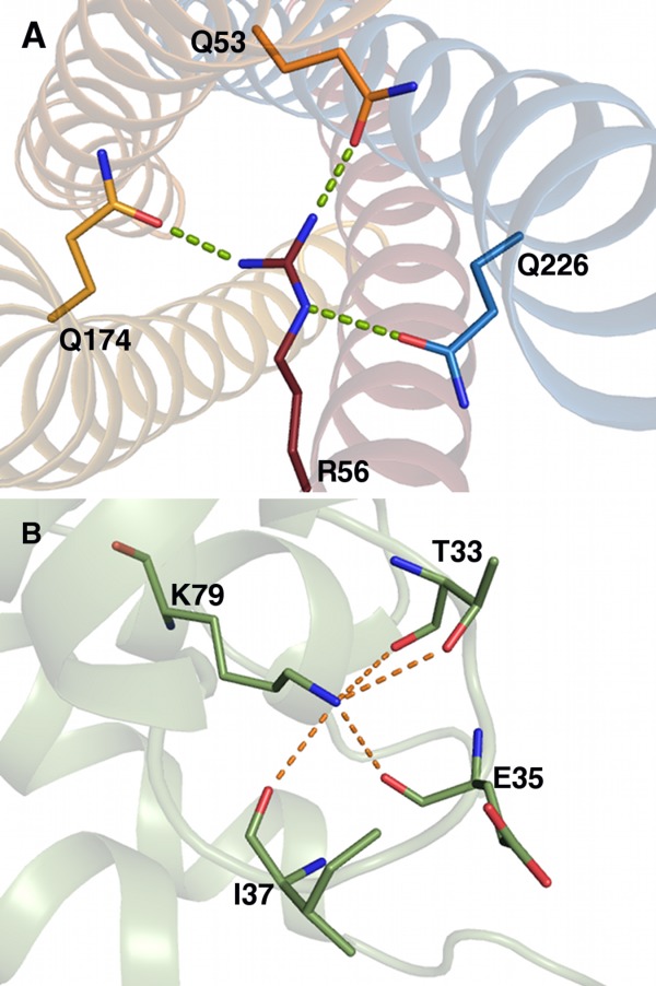
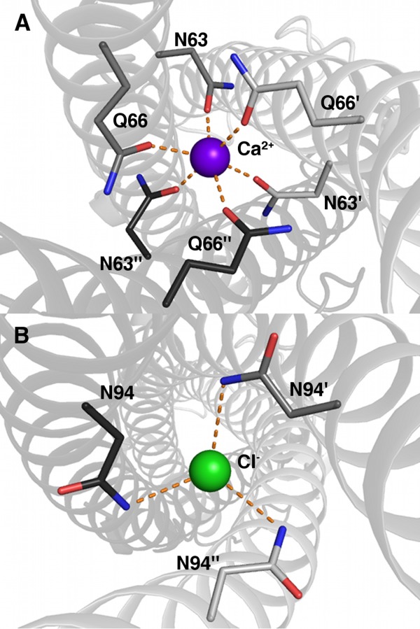
Ion-dipole interactions in biological macromolecules are formed between atomic or molecular ions and neutral protein dipolar groups through either hydrogen bond or coordination. Since their discovery 30 years ago, these interactions have proven to be a frequent occurrence in protein structures, appearing in everything from transporters and ion channels to enzyme active sites to protein-protein interfaces. However, their significance and roles in protein functions are largely underappreciated. We performed PDB data mining to identify a sampling of proteins that possess these interactions. In this review, we will define the ion-dipole interaction and discuss several prominent examples of their functional roles in nature.

Keywords: ABC transport receptors; AP180; IMP dehydrogenase; Ion-dipole interaction; SNARE; ion channel; phage tail proteins; serine protease.


Figures

Figure 1
Figure 1
Salmonella typhimurium SBP-sulfate complex X-ray structure (PDB ID 1sbp). (A) SBP sulfate-binding site. Dashed lines represent ion–dipole interactions via hydrogen bonds, and cooperative hydrogen bonds are colored purple. Carbons are colored green; nitrogen, blue; oxygen, red; sulfur, gold. (B) Helix N-termini in the sulfate-binding site, each providing a main chain NH dipole.
Figure 2
Figure 2
Streptomyces lividans KcsA potassium channel X-ray structure (PDB ID 1bl8) potassium site S4. Dashed lines represent ion–dipole interactions via coordination, and cooperative hydrogen bonds are colored green. Oxygens are colored red; nitrogens, blue; and potassium, purple. Different chain carbons are colored as different shades of orange and labeled with primes.
Figure 3
Figure 3
Porcine pancreatic elastase complexed with a peptidyl inhibitor FR130180 neutron structure (PDB ID 3hgn) oxyanion hole. Hydrogenated protein was crystallized in a precipitant solution prepared in D2O, which caused the exchangeable hydrogens to be replaced with deuteriums prior to neutron diffraction. The Ser and Gly stabilize the charged catalytic intermediate. Dashed lines represent ion–dipole interactions. Protein carbons are colored tan; FR130180 carbons, green; nitrogen, dark blue; oxygen, red; hydrogens, white; and deuteriums, light cyan.
Figure 4
Figure 4
Vibrio cholerae inosine 5′-monophosphate (IMP) dehydrogenase X-ray structure. (A) IMP dehydrogenase potassium-binding site (PDB ID 4qne). Dashed lines represent ion dipole via coordination. Protein carbons are colored pink; symmetry-related carbons, gray; nitrogen, blue; oxygen, red; sulfur, yellow; phosphorus, orange; and potassium, purple. Symmetry-related residues are labeled with primes. (B) Potassium-bound superposed on apo-IMP dehydrogenase (colored cyan, PDB ID 4qq3). Areas structured upon K+ binding noted with red circles.
Figure 5
Figure 5
(A) Rattus norvegicus SNARE complex X-ray structure (PDB ID 1sfc). The synaptobrevin Arg interacts with Glns from syntaxin and the N- and C-terminal segments of synaptosome-associated protein of relative molecular mass 25 kDa (SNAP 25). Dashed lines represent ion–dipoles. Syntaxin carbons are colored dark blue; synaptobrevin carbons, red; SNAP 25 carbons, orange; nitrogen, blue; and oxygen, red. (B) Drosophila melanogaster AP180 X-ray structure (PDB ID 1sfc). The Lys stabilizes the active site loop, maintaining an active conformation. Carbons are colored green; nitrogen, blue; and oxygen, red.
Figure 6
Figure 6
Enterobacteria phage P22 tail needle protein gp26 X-ray structure (PDB ID 3c9i) showing ion–dipole interactions by way of calcium (A) and chloride (B) coordination. Dashed lines represent ion–dipole interactions. (A) Calcium-binding site. Oxygens are colored red; nitrogens, blue; and calcium, purple. Different chain carbons are colored as different shades of gray and labeled with primes. (B) Chlorine-binding site. The protein is labeled and colored as in (A), and chlorine is colored green.

References

    1. Pflugrath JW, Quiocho FA. Sulphate sequestered in the sulphate-binding protein of Salmonella typhimurium is bound solely by hydrogen bonds. Nature. 1985;314:21–27. - PubMed
    1. Cannon R, Pettitt BM, McCammon JA. Sulfate anion in water: model structural, thermodynamic, and dynamic properties. J Phys Chem. 1994;98:6225–6230.
    1. Kelly CP, Cramer CJ, Truhlar DG. Aqueous solvation free energies of ions and ion-water clusters based on an accurate value for the absolute aqueous solvation free energy of the proton. J Phys Chem B. 2006;110:16066–16081. - PubMed
    1. Marcus Y. Ion solvation. Chichester, UK: Wiley; 1985.
    1. Honig B, Yang AS. Free energy balance in protein folding. Adv Prot Chem. 1995;46:27–58. - PubMed

Publication types

LinkOut - more resources