. 2014 Dec 1;9(6):695-704.
doi: 10.1016/j.nantod.2014.10.003.

A Multilayered Cell Culture Model for Transport Study in Solid Tumors: Evaluation of Tissue Penetration of Polyethyleneimine Based Cationic Micelles

Affiliations

A Multilayered Cell Culture Model for Transport Study in Solid Tumors: Evaluation of Tissue Penetration of Polyethyleneimine Based Cationic Micelles

Seiji Miura et al. Nano Today. .

Abstract

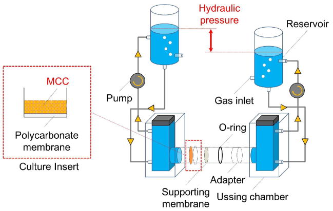
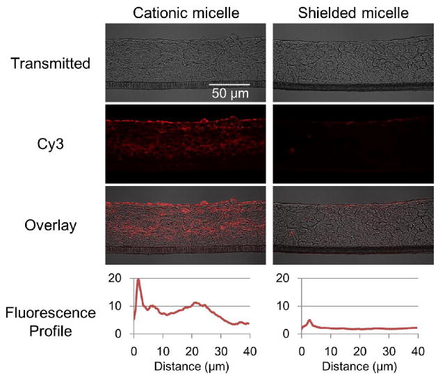
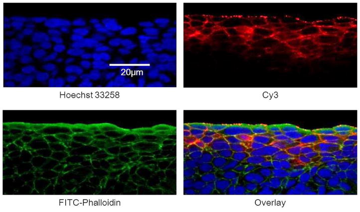
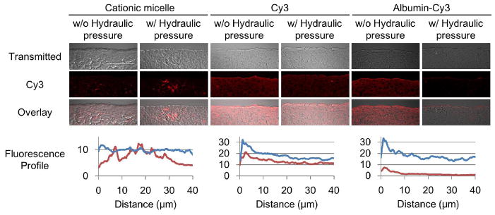
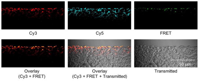
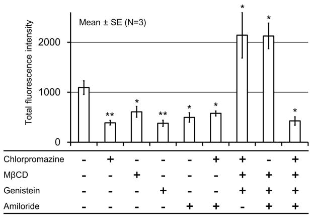
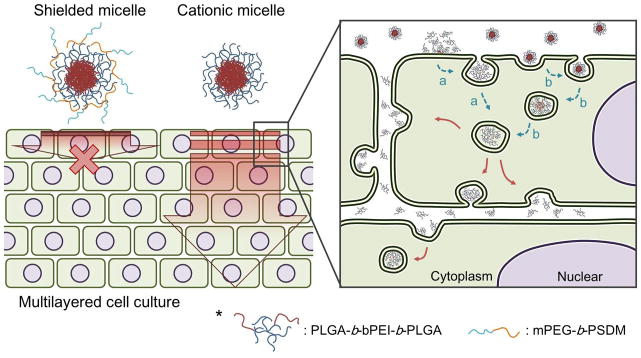
Limited drug distribution is partially responsible for the efficacy gap between preclinical and clinical studies of nano-sized drug carriers for cancer therapy. In this study, we examined the transport behavior of cationic micelles formed from a triblock copolymer of poly(D,L-lactide-co-glycolide)-block-branched polyethyleneimine-block-poly(D,L-lactide-co-glycolide) using a unique in vitro tumor model composed of a multilayered cell culture (MCC) and an Ussing chamber system. The Cy3-labeled cationic micelles showed remarkable Cy3 distribution in the MCC whereas charge-shielded micelles with a poly(ethylene glycol) surface accumulated on the surface of the MCC. Penetration occurred against convectional flow caused by a hydraulic pressure gradient. The study using fluorescence resonance energy transfer (FRET) showed that the cationic micelles dissociate at the interface between the culture media and the MCC or possibly inside of the first-layer cells and penetrates into the MCC as unimers. The penetration and distribution were energy-dependent and suppressed by various endocytic inhibitors. These suggest that cationic unimers mainly utilized clathrin-mediated endocytosis and macropinocytosis for cellular entry and a significant fraction were exocytosed by an unknown mechanism.

Keywords: cationic surface; endocytosis; intratumoral distribution; micelle; multilayered cell culture; penetration.


Figures

Figure 1
Figure 1
Modified Ussing chamber system. The multilayered cell culture (MCC) was secured into the Ussing chamber by an O-ring and an adapter and the top of the MCC was faced to donor chamber. Each chamber was connected to the reservoir with silicone tubing. The culture medium in each chamber was circulated by a pump. The supporting membrane with a mesh size of 40 μm was used to prevent the separation of the MCC from the culture insert by pressure gradients. The whole system was maintained at 37 °C by a heat block (not shown in the figure).
Figure 2
Figure 2
Distribution of cationic micelles and shielded micelles in the MCC. MCCs were exposed to the cationic and shielded micelles labeled with Cy3 (red) at 37 °C for one hour. Cryo-sections were prepared and imaged by confocal microscopy. Fluorescence was presented as an intensity profile over distance from the MCC surface using ImageJ [66]. The images are representative and the profiles are average of data set from three different cross sections.
Figure 3
Figure 3
Localization of Cy3 from cationic micelles in the MCC. The MCC was exposed to the cationic micelles labeled with Cy3 (red) at 37 °C for one hour. Cryo-sections were prepared and stained with Hoechst 33258 for the nucleus (blue), FITC-phalloidin for the cytoskeleton (green).
Figure 4
Figure 4
Influence of hydraulic pressure on the penetration of the Cy3 labeled micelles and albumins. The MCCs were exposed to the cationic micelles conjugated with Cy3 (red) and control molecules: free Cy3 (1 μg/mL) and Cy3 conjugated albumins (2.5 mg/mL) at 37 °C for one hour with or without a hydraulic pressure gradient. Cryo-sections were prepared and then imaged by confocal microscopy. Fluorescence was presented as an intensity profile over distance using ImageJ [66]. The profiles for distribution without the hydraulic pressure gradient are shown in blue, and with the hydraulic pressure gradient in red. Scale bar = 50 μm. The images are representative and the profiles are average of data set from three different cross sections.
Figure 5
Figure 5
The penetration of the cationic micelles in the MCC. The MCC was exposed to the FRET micelles at 37 °C for one hour. The emission of Cy5 (λEm= >650 nm) was detected as a FRET signal using the excitation wave length of Cy3 (λEx=543 nm). The images are representative.
Figure 6
Figure 6
Inhibition of unimer penetration by tannic acid and low temperature. The MCC was exposed to cationic micelles conjugated with Cy3 (red) for one hour at 37 or at 4 °C with or without pretreatment of tannic acid. Cryo-sections were prepared and then imaged by confocal microscopy. Fluorescence was presented as an intensity profile over distance using ImageJ [66]. Unimer penetration profile at 37 °C is shown in blue, and the Cy3 profiles by treating the MCC with tannic acid and a low temperature (4 °C) are in red. The images are representative and the profiles are average of data set from three different cross sections.
Figure 7
Figure 7
Inhibition of cationic unimer penetration by various inhibitors. The MCC was exposed to the cationic micelles labeled with Cy3 (red) at 37 °C for one hour with or without pretreatment of inhibitiors. Cryo-sections were prepared and then imaged by confocal microscopy. Total fluorescence intensity is presented as the area under the curve of fluorescence profiles shown in Supplementary Fig. S5. *p<0.05, **p<0.01
Figure 8
Figure 8
Proposed penetration mechanism of cationic micelles through a multilayered cell culture. The cationic micelles penetrate through a multilayered cell culture as unimers. Dissociation of micelles is possibly caused on a surface of cell membranes (a) or inside of first layer cells.

References

    1. Wang AZ, Langer R, Farokhzad OC. Annu Rev Med. 2012;63:185–198. - PubMed
    1. Zhang L, Gu FX, Chan JM, Wang AZ, Langer RS, Farokhzad OC. Clin Pharmacol Ther. 2008;83:761–769. - PubMed
    1. Stirland DL, Nichols JW, Miura S, Bae YH. J Control Release. 2013;172:1045–1064. - PMC - PubMed
    1. Lammers T, Kiessling F, Hennink WE, Storm G. J Control Release. 2012;161:175–187. - PubMed
    1. Adams DJ. Trends Pharmacol Sci. 2012;33:173–180. - PMC - PubMed