

Recent advances in 3D printing of biomaterials
- PMID: 25866560
- PMCID: PMC4392469
- DOI: 10.1186/s13036-015-0001-4
Recent advances in 3D printing of biomaterials
Abstract
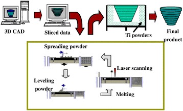
3D Printing promises to produce complex biomedical devices according to computer design using patient-specific anatomical data. Since its initial use as pre-surgical visualization models and tooling molds, 3D Printing has slowly evolved to create one-of-a-kind devices, implants, scaffolds for tissue engineering, diagnostic platforms, and drug delivery systems. Fueled by the recent explosion in public interest and access to affordable printers, there is renewed interest to combine stem cells with custom 3D scaffolds for personalized regenerative medicine. Before 3D Printing can be used routinely for the regeneration of complex tissues (e.g. bone, cartilage, muscles, vessels, nerves in the craniomaxillofacial complex), and complex organs with intricate 3D microarchitecture (e.g. liver, lymphoid organs), several technological limitations must be addressed. In this review, the major materials and technology advances within the last five years for each of the common 3D Printing technologies (Three Dimensional Printing, Fused Deposition Modeling, Selective Laser Sintering, Stereolithography, and 3D Plotting/Direct-Write/Bioprinting) are described. Examples are highlighted to illustrate progress of each technology in tissue engineering, and key limitations are identified to motivate future research and advance this fascinating field of advanced manufacturing.
Keywords: 3D Printing; 3D plotting; Bioprinting; Computer-aided tissue engineering; Fused deposition modeling; Selective laser sintering; Stereolithography.
Figures


References
-
- Karande TS, Ong JL, Agrawal CM. Diffusion in musculoskeletal tissue engineering scaffolds: design issues related to porosity, permeability, architecture, and nutrient mixing. Ann Biomed Eng. 2004;32:1728–43. - PubMed
-
- Hollister SJ. Porous scaffold design for tissue engineering. Nat Mater. 2005;4:518–24. - PubMed
-
- Stevens MM, George JH. Exploring and engineering the cell surface interface. Science. 2005;310:1135–8. - PubMed
-
- Winder J, Bibb R. Medical rapid prototyping technologies: state of the art and current limitations for application in oral and maxillofacial surgery. J Oral Maxillofac Surg. 2005;63:1006–15. - PubMed
-
- Colin A, Boire J-Y. A novel tool for rapid prototyping and development of simple 3D medical image processing applications on PCs. Comput Methods Programs Biomed. 1997;53:87–92. - PubMed
LinkOut - more resources
Full Text Sources
Other Literature Sources
Medical
