Review
. 2015 Apr-Jun;8(2):120-8.

Considerations regarding current diagnosis and prognosis of hepatocellular carcinoma

Affiliations
Review

Considerations regarding current diagnosis and prognosis of hepatocellular carcinoma

C G Cristea et al. J Med Life. 2015 Apr-Jun.

Abstract

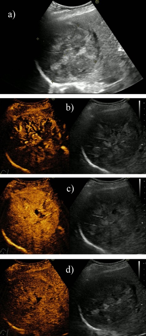
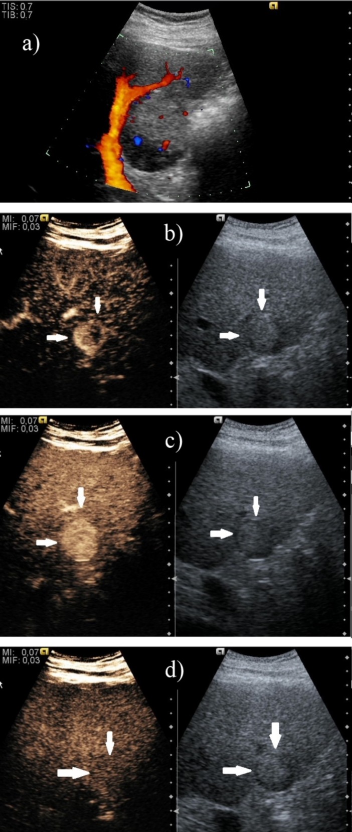
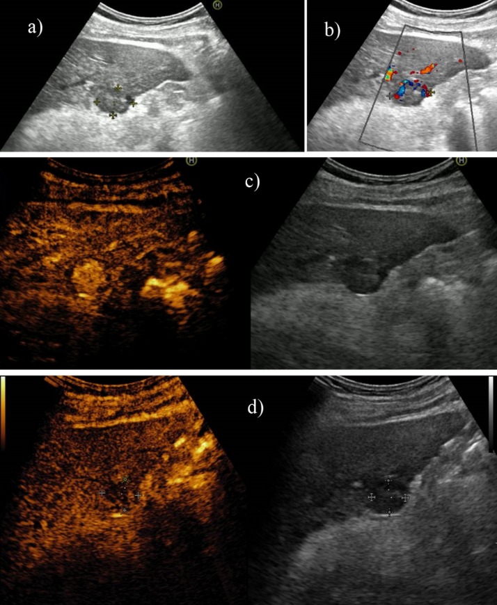
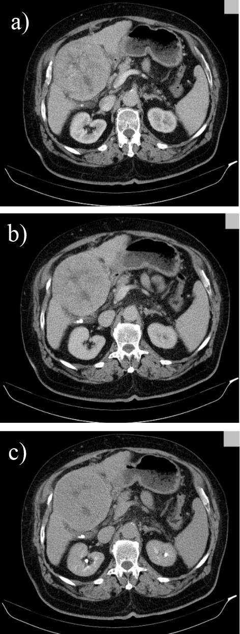
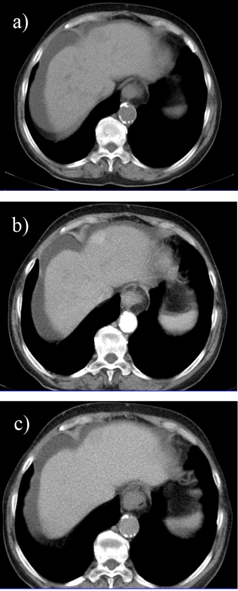
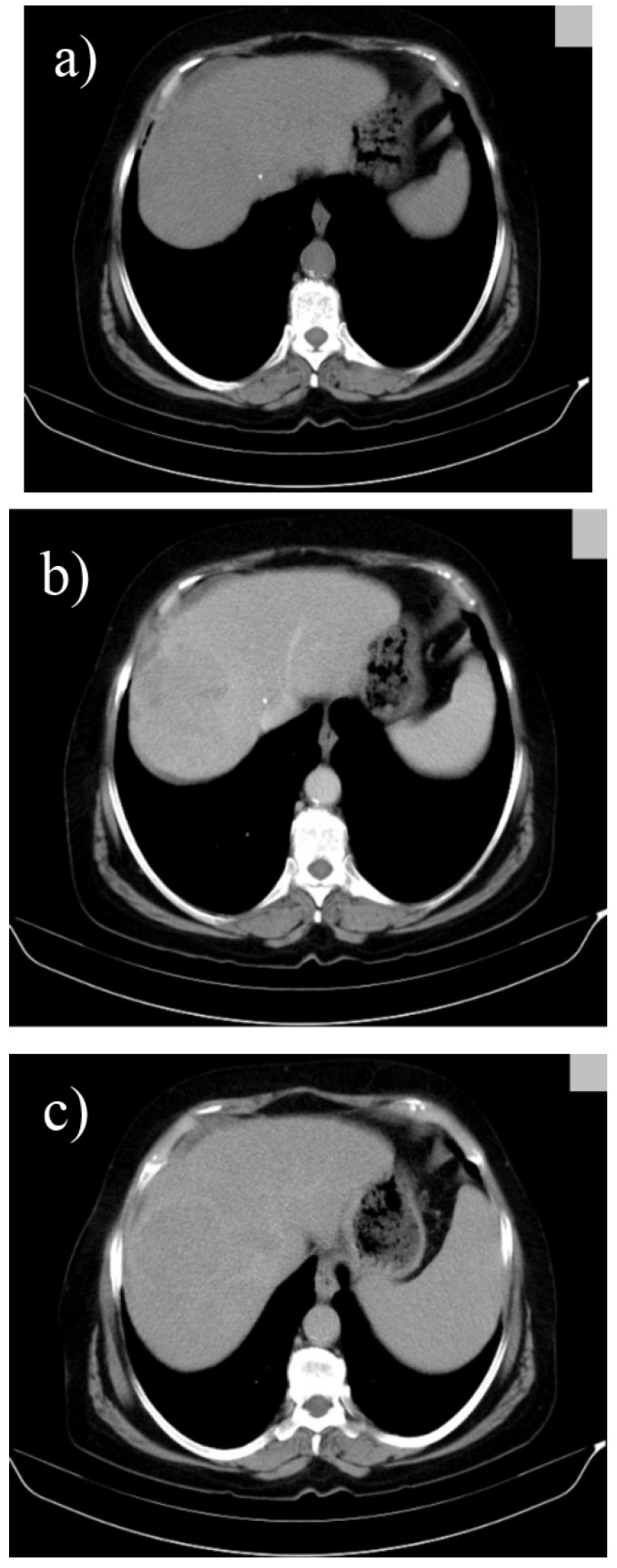
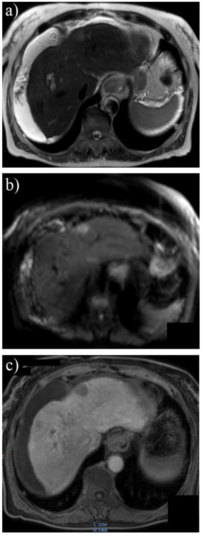
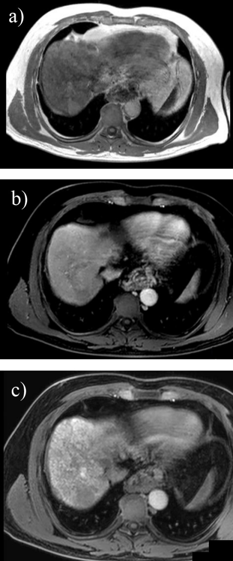
Hepatocellular carcinoma is a major health issue, ranked the fifth most common tumor and currently being responsible for a third of the cancer-related deaths globally, with an ever-increasing number of fatalities. Current advances in contrast-enhanced imaging techniques such as contrast-enhanced ultrasonography, multi-detector computed tomography and diffusion-weighted magnetic resonance imaging are improving the rate of hepatocellular carcinoma diagnosis. Contrast-enhanced ultrasonography has widely become the first choice in liver tumor assessment, as it is faster, simpler and safer than other forms of diagnostic imaging. On the other hand, cross sectional computed tomography is frequently employed when a hepatic formation is suspected of malignancy and allows a more accurate characterization of lesions through multiphasic multi-detector computed tomography technology. Diffusion weighted magnetic resonance imaging represents another addition to the wide range of diagnostic and prognostic techniques available for patients with hepatocellular carcinoma and is currently regarded as one of the best tools for the characterization of these lesions. Furthermore, groundbreaking biomarkers for hepatocellular carcinoma are being discovered, although alpha-fetoprotein remains one of the most frequently used serum test in the early stages. Nonetheless, further advances are required for the detection of small liver carcinomas.

Keywords: alpha-fetoprotein; contrast-enhanced ultrasonography; diffusion weighted MRI; hepatocellular carcinoma; multi-detector CT.


Figures

Fig. 1
Fig. 1
Large HCC (a), which presents on CEUS chaotic intratumoral vascularization (b), incomplete contrast filling during the arterial phase due to areas of tumor necrosis (c), as well as slow and incomplete washout after approximately 5 minutes of examination (d)
Fig. 2
Fig. 2
Well-differentiated hepatocellular carcinoma with discreet vascularization on Doppler ultrasound (a), but with intense and heterogeneous hyperenhancement during the arterial phase (b, c) and without washout during the late phase (d)
Fig. 3
Fig. 3
In a 56-year-old patient, standard ultrasound showed a small nodule of 1,8 cm in the hepatic segment V (a) With “basket pattern” type vascularization on Doppler ultrasound (b) In the arterial phase, the nodule fills with contrast faster and more intense compared to the hepatic parenchyma (c) and presents washout in the late phase (d)
Fig. 4
Fig. 4
Arterial (a), portal (b) and late phase (c) 16 slices CT in a cirrhotic patient revealing a well-differentiated hepatocellular carcinoma in segment IV with extracapsular extension, heterogenous contrast uptake and central necrosis
Fig. 5
Fig. 5
Triphasic multislice CT in a cirrhotic patient showing a moderatly differented small, subcapsular HCC in segment IV with slight hypoattenuation in the precontrast phase (a), with arterial enhancement (b) and washout in the portal phase (c);
Fig. 6
Fig. 6
Triphasic multislice CT in a cirrhotic patient showing a well differented HCC in the right lobe with the same density as liver parenchyma in precontrast phase (a), with discrete low density during portal phase (b) and low density with hiperattenuating capsule in the late phase (c)
Fig. 7
Fig. 7
3T MRI in a patient with small HCC in segment II with high signal intensity in T2 weighted (a) and diffusion (b) sequences and decreased signal intensity at 30 minutes after contrast enhancement with hepatospecific paramagnetic gadolinium based contrast agent (c)
Fig. 8
Fig. 8
3T MRI in a cirrhotic patient using Gd-EOB-DTPA and revealing a small nodule in segment VII with high signal in axial T1 in phase (a), low signal in dynamic postcontrast (b) and at 30 minutes in hepatobilliary phase (c) suggesting the diagnosis of HCC

References

    1. Marrero JA, Feng Z, Wang Y, et al. Alpha-fetoprotein, des-gamma carboxyprothrombin, and lectin-bound alpha-fetoprotein in early hepatocellular carcinoma. Gastroenterology. 2009;137:110–118. - PMC - PubMed
    1. Ariff B, Lloyd CR, Khan S, et al. Imaging of liver cancer. World Journal of Gastroenterology: WJG. 2009;15:1289–1300. - PMC - PubMed
    1. Eusebi P. Diagnostic accuracy measures. Cerebrovascular Diseases. 2013;36:267–272. - PubMed
    1. Jang HJ, Yu H, Kim TK. Contrast-enhanced ultrasound in the detection and characterization of liver tumors. Cancer imaging: the official publication of the International Cancer Imaging Society. 2009;9:96–103. - PMC - PubMed
    1. Danila M, Sporea I, Sirli R, et al. The role of contrast enhanced ultrasound (CEUS) in the assessment of liver nodules in patients with cirrhosis. Medical Ultrasonography. 2010;12:145–149. - PubMed

Publication types

MeSH terms

Substances

LinkOut - more resources