Review
. 2015:2015:854024.
doi: 10.1155/2015/854024. Epub 2015 Mar 18.

Updates and knowledge gaps in cholesteatoma research

Affiliations
Review

Updates and knowledge gaps in cholesteatoma research

Chin-Lung Kuo et al. Biomed Res Int. 2015.

Abstract

The existence of acquired cholesteatoma has been recognized for more than three centuries; however, the nature of the disorder has yet to be determined. Without timely detection and intervention, cholesteatomas can become dangerously large and invade intratemporal structures, resulting in numerous intra- and extracranial complications. Due to its aggressive growth, invasive nature, and the potentially fatal consequences of intracranial complications, acquired cholesteatoma remains a cause of morbidity and death for those who lack access to advanced medical care. Currently, no viable nonsurgical therapies are available. Developing an effective management strategy for this disorder will require a comprehensive understanding of past progress and recent advances. This paper presents a brief review of background issues related to acquired middle ear cholesteatoma and deals with practical considerations regarding the history and etymology of the disorder. We also consider issues related to the classification, epidemiology, histopathology, clinical presentation, and complications of acquired cholesteatoma and examine current diagnosis and management strategies in detail.


Figures

Figure 1
Figure 1
(a) Congenital cholesteatoma, anterosuperior quadrant of eardrum (left ear). A white mass is located behind an intact eardrum without prior otitis media or history of otologic procedures. (b) Dissected round mass with whitish pearly appearance.
Figure 2
Figure 2
(a) Acquired cholesteatoma of attic in the left ear (arrow). Accumulation of debris within an attic retraction pocket led to gradual expansion of cholesteatoma. Grommet insertion revealed poor ventilation function in the middle ear (arrow head). (b) Dissected friable cholesteatoma with a thin pearly-white greasy-looking wall containing pultaceous substance.
Figure 3
Figure 3
Histopathology of cholesteatoma showing a central mass of keratin (cystic content) surrounded by a thin layer of stratified squamous epithelium (matrix) and fibrous tissue with inflammatory infiltrate (perimatrix) (H and E, ×40).
Figure 4
Figure 4
Schematic representation of the paracrine and autocrine interactions between matrix keratinocytes and perimatrix fibroblasts. Keratinocytes release proinflammatory cytokines (e.g., IL-1α, IL-1β, IL-6, PTHrP, and IL-8), which subsequently induce fibroblasts to secrete several cytokines (e.g., KGF, GM-CSF, EGF, TNF-α, PDGF, and TGF-α). These fibroblast-derived cytokines in turn induce the differentiation, proliferation, and migration of matrix keratinocytes. In addition, the TGF-α and TGF-β are upregulated in an autocrine loop, regulating keratinocyte proliferation and differentiation. EGF: epidermal growth factor; GM-CSF: granulocyte-macrophage colony stimulating factor; IL: interleukin; KGF: keratinocytes growth factor; PDGF: platelet-derived growth factor; PTHrP: parathyroid-hormone-related protein; TGF: transforming growth factor; TNF-α: tumor necrosis factor alpha.
Figure 5
Figure 5
Cholesteatoma can be easily overlooked when hidden over the outer attic wall by various substances such as crust (a), debris (c), and granulation tissue (e). Complete removal of these substances may prevent misdiagnosis of cholesteatoma deep within the middle ear cleft ((b), (d), and (f)).
Figure 6
Figure 6
Attic retraction pocket in the left ear (white arrow) with atelectatic Prussak's space (red circle) and eroded scutum (yellow arrow).
Figure 7
Figure 7
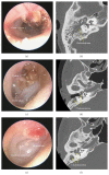
(a) Axial and (b) coronal CT scans of right temporal bone showing cholesteatoma with cochlear fistula (arrow). (c) Intraoperative findings confirmed the fistula with pulsatile fluid. The arrow head indicates light reflex in the fluid.
Figure 8
Figure 8
(a) Normal contour of the external ear canal in the left ear. (b) Traditional canal wall down mastoidectomy involves removing the posterior canal wall, which results in the formation of an open cavity. Regular ear cleaning is required to remove accumulated debris and control infection.

References

    1. Isaacson G. Diagnosis of pediatric cholesteatoma. Pediatrics. 2007;120(3):603–608. doi: 10.1542/peds.2007-0120. - DOI - PubMed
    1. Semaan M. T., Megerian C. A. The pathophysiology of cholesteatoma. Otolaryngologic Clinics of North America. 2006;39(6):1143–1159. doi: 10.1016/j.otc.2006.08.003. - DOI - PubMed
    1. Dornelles C., da Costa S. S., Meurer L., Schweiger C. Some considerations about acquired adult and pediatric cholesteatomas. Revista Brasileira de Otorrinolaringologia. 2005;71(4):536–546. doi: 10.1590/s0034-72992005000400023. - DOI - PMC - PubMed
    1. Robinson J. M. Cholesteatoma: skin in the wrong place. Journal of the Royal Society of Medicine. 1997;90(2):93–96. - PMC - PubMed
    1. Gray J. D. The chronic ear. The treatment of cholesteatoma in children. Proceedings of the Royal Society of Medicine. 1964;57:769–771. - PMC - PubMed

MeSH terms

LinkOut - more resources