. 2015 May;125(5):2007-20.
doi: 10.1172/JCI78124. Epub 2015 Apr 13.

Ash1l controls quiescence and self-renewal potential in hematopoietic stem cells

Ash1l controls quiescence and self-renewal potential in hematopoietic stem cells

Morgan Jones et al. J Clin Invest. 2015 May.

Abstract

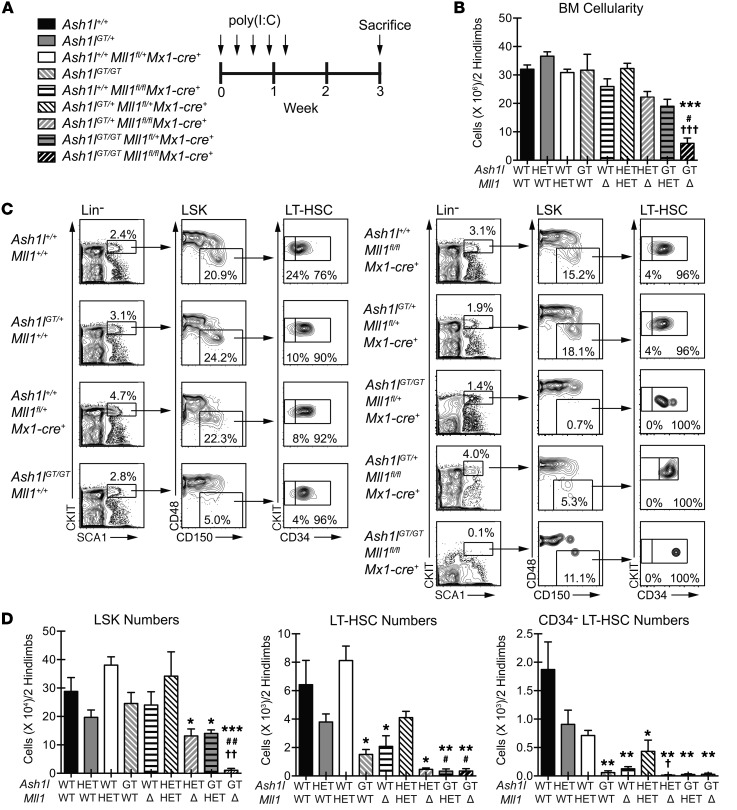
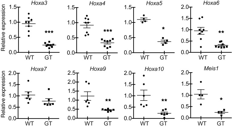
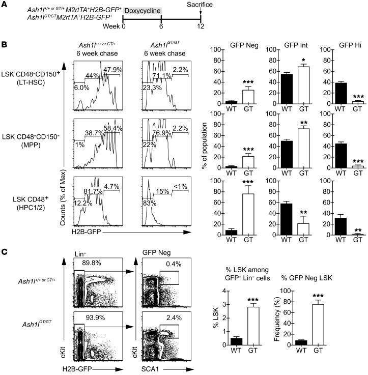
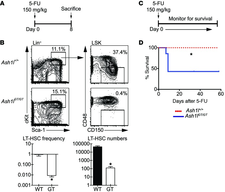
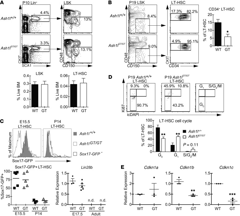
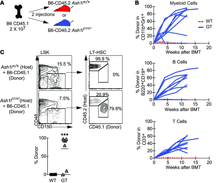
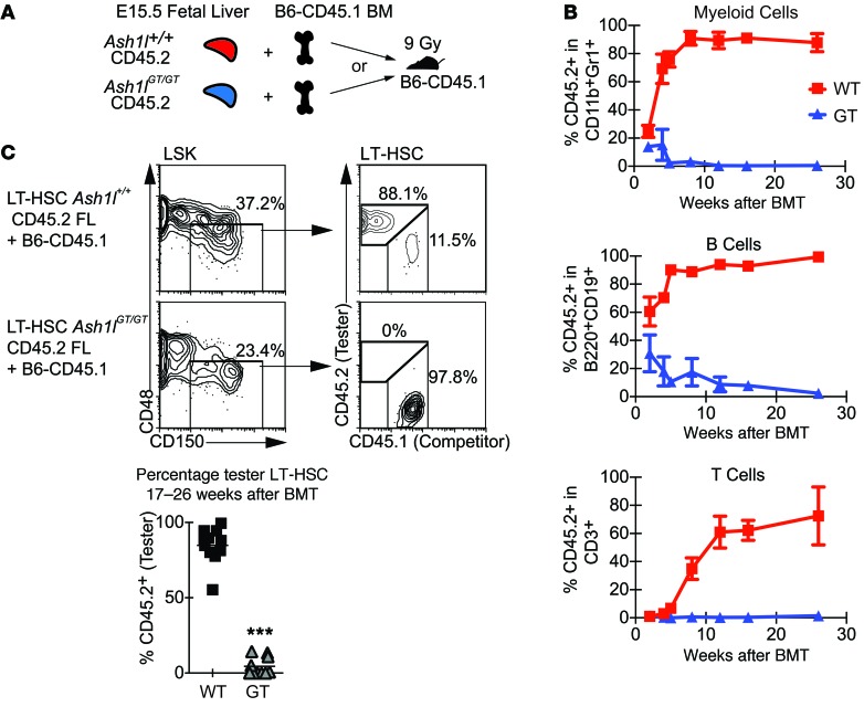
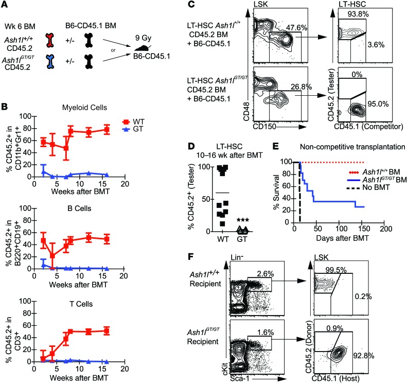
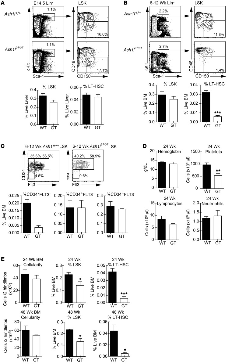
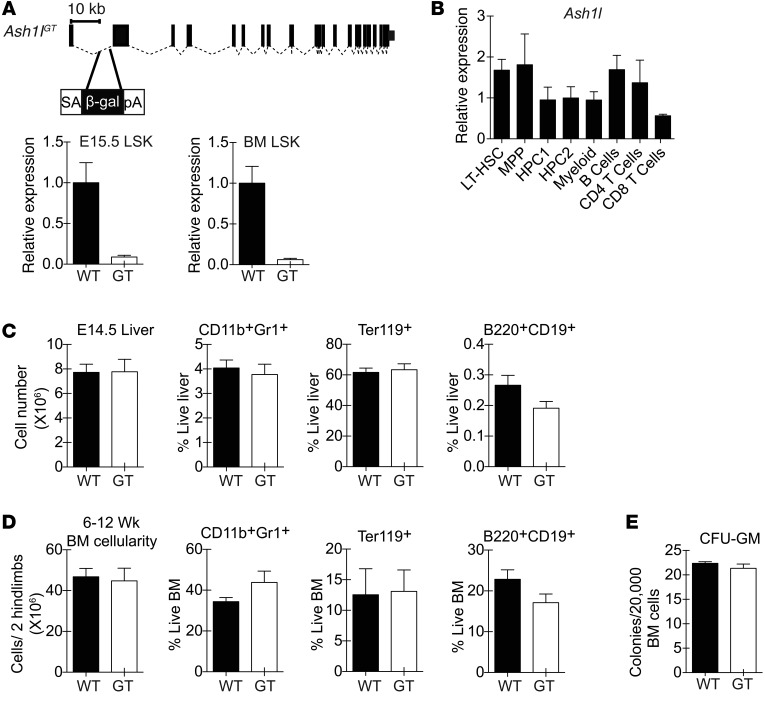
Rapidly cycling fetal and neonatal hematopoietic stem cells (HSCs) generate a pool of quiescent adult HSCs after establishing hematopoiesis in the bone marrow. We report an essential role for the trithorax group gene absent, small, or homeotic 1-like (Ash1l) at this developmental transition. Emergence and expansion of Ash1l-deficient fetal/neonatal HSCs were preserved; however, in young adult animals, HSCs were profoundly depleted. Ash1l-deficient adult HSCs had markedly decreased quiescence and reduced cyclin-dependent kinase inhibitor 1b/c (Cdkn1b/1c) expression and failed to establish long-term trilineage bone marrow hematopoiesis after transplantation to irradiated recipients. Wild-type HSCs could efficiently engraft when transferred to unirradiated, Ash1l-deficient recipients, indicating increased availability of functional HSC niches in these mice. Ash1l deficiency also decreased expression of multiple Hox genes in hematopoietic progenitors. Ash1l cooperated functionally with mixed-lineage leukemia 1 (Mll1), as combined loss of Ash1l and Mll1, but not isolated Ash1l or Mll1 deficiency, induced overt hematopoietic failure. Our results uncover a trithorax group gene network that controls quiescence, niche occupancy, and self-renewal potential in adult HSCs.


Figures

Figure 10
Figure 10. Combined Ash1l and Mll1 deficiency induces overt hematopoietic failure and profound depletion of LT-HSCs and LSK progenitors.
(A) Experimental strategy: mice of indicated genotypes were injected with poly(I:C) (20 μg every 2 days 5 times). (B) Reduced BM cellularity in Ash1lGT/GT Mll1fl/flMx1-Cre+mice (≥2 mice per genotype; mean ± SEM). (C) Flow cytometric analysis showing severe reduction in CD34 LT-HSCs, LT-HSCs, and LSK progenitors in Ash1lGT/GT Mll1fl/flMx1-Cre+mice and reduced CD34LT-HSC and LT-HSC frequency with cumulative inactivation of Ash1l and Mll1 alleles. The representative plot for Ash1lGT/+ Mll1fl/flMx1-Cre+ BM is derived from a separate experiment, thus gating definitions differ from the other samples. (D) LSK and LT-HSC numbers, reflecting hematopoietic failure in Ash1lGT/GT Mll1fl/flMx1-Cre+ mice and LT-HSC sensitivity to loss of Ash1l and Mll1 alleles (≥2 mice per genotype; mean ± SEM), and CD34 LT-HSC numbers in 2 hind legs, showing high sensitivity of this population to regulation by TrxG members. *P < 0.05, **P < 0.01, ***P < 0.001, compared with wild type; #P < 0.05, ##P < 0.01, compared with Ash1lGT/GT; P < 0.05, ††P < 0.01, †††P < 0.001, compared with induced Mllfl/fl Mx1-Cre+, t test.
Figure 9
Figure 9. Ash1l regulates expression of Hox and Hox-related genes in hematopoietic progenitors.
Relative abundance of Hoxa and Meis1 normalized to Hprt1 transcripts in sort-purified adult Ash1lGT/GT LSK progenitors, as assessed by qRT-PCR (triplicate analysis of 4 to 8 individual biological samples per genotype, mean ± SEM; *P < 0.05, **P < 0.01, ***P < 0.001, t test).
Figure 8
Figure 8. In vivo proliferation history shows decreased quiescence of Ash1l-deficient HSCs but increased persistence of proliferating downstream progenitors.
(A) Experimental strategy: Ash1l+/+ or Ash1lGT/GT mice with M2rtTA and H2B-GFP transgenes were maintained on doxycycline for 6 weeks to label hematopoietic cells with GFP. GFP dilution was monitored by flow cytometry after a 6-week chase period. (B) Flow cytometric analysis after chase, showing significantly increased GFP dilution in Ash1lGT/GT LSK CD150+CD48 cells (containing LT-HSCs), LSK CD150CD48 (MPP), and LSK CD48+ progenitors (HPC1/2) compared with controls, consistent with increased cell division (≥6 mice per genotype; mean ± SEM). (C) Flow cytometric analysis, demonstrating that the H2B-GFP–negative fraction of Lin cells was enriched for primitive LSK progenitors in Ash1lGT/GT versus Ash1l+/+ mice (≥ 4 mice per genotype; mean ± SEM). *P < 0.05, **P < 0.01, ***P < 0.001, t test.
Figure 7
Figure 7. Ash1lGT/GT mice are more sensitive to 5-FU challenge than wild-type mice.
(A) Experimental strategy: mice of indicated genotypes were injected with 150 mg/kg 5-FU and sacrificed 8 days later. (B) Flow cytometric analysis of LT-HSCs (CD150+CD48LSK cells), showing 2-log reduction in frequency and 2.5-log reduction in LT-HSC numbers in Ash1lGT/GT mice as compared with control mice after 5-FU exposure (n ≥ 4 mice per genotype; mean ± SEM, *P < 0.05, t test). (C) Experimental strategy: mice were injected with 150 mg/kg 5-FU and monitored for survival. (D) Survival of mice after 5-FU challenge (7 mice per genotype, representative of 2 experiments, *P < 0.05, log-rank Mantel-Cox test).
Figure 6
Figure 6. Ash1lGT/GT LT-HSCs home to the BM but fail to establish normal quiescence, despite appropriate extinction of the fetal HSC program.
(A) Flow cytometric analysis of P10 BM, showing comparable frequencies of phenotypically defined LT-HSCs (CD150+CD48LSK) in Ash1lGT/GT and Ash1l+/+ littermates (≥9 mice per genotype; mean ± SEM). (B) Decreased percentage of quiescent CD34 cells in Ash1lGT/GT LT-HSCs (P19) (4 mice per genotype; mean ± SEM). (C) Sox17-GFP expression in E15.5 fetal liver and P14 BM LT-HSCs (CD150+CD48LSK cells). Sox17-GFP was present in fetal LT-HSCs but extinguished in wild-type and Ash1lGT/GT P14 BM (n ≥ 3 per genotype, 4 experiments; symbols show individual mice and the bars show the mean). qRT-PCR analysis shows similar Lin28b gene expression in Ash1lGT/GT and Ash1l+/+ fetal LSK progenitors, with loss of expression in adult progenitors of both genotypes (n = 3–5 per group; mean ± SEM) (n.d., not detectable). (D) Flow cytometry plots (Ki67 vs. DAPI) showing decreased G0 (quiescent fraction) and increased distribution into G1 and S/G2/M phases of the cell cycle in P19 Ash1lGT/GT LT-HSCs (5 mice per genotype; mean ± SEM). (E) Reduced Cdkn1b and Cdkn1c expression relative to Hprt1 in P10 Ash1lGT/GT LSK progenitors (qRT-PCR, n = 3 per group, mean ± SEM). *P < 0.05, **P < 0.01, ***P < 0.001, t test.
Figure 5
Figure 5. Ash1l deficiency allows engraftment of wild-type HSCs in the absence of myeloablation.
(A) Experimental strategy: B6-CD45.2 Ash1l+/+ or Ash1lGT/GT mice received 2 × 10⁷ B6-CD45.1 or B6-GFP BM cells (2 doses 1 week apart i.v.), without prior irradiation. (B) Analysis of peripheral blood, showing myeloid, B, and T cell output from CD45.1+ donor BM in 7 of 8 Ash1lGT/GT recipients versus 0 of 6 Ash1l+/+ recipients (data from 3 experiments). Lines represent individual recipients. Reconstitution was sustained for ≥12 weeks. (C) Flow cytometric analysis of LT-HSCs 12 to 21 weeks after infusion, showing donor LT-HSC engraftment in 7 of 8 Ash1lGT/GT recipients versus 0 of 6 wild-type recipients (pooled from 3 experiments, with horizontal bars showing the mean; ***P < 0.001, t test).
Figure 4
Figure 4. Ash1l-deficient fetal liver cells do not sustain long-term BM reconstitution after competitive transplantation.
(A) Experimental strategy: Ash1l+/+or Ash1lGT/GT B6-CD45.2 E15.5 fetal liver was mixed with wild-type B6-CD45.1 competitor BM (1:1 ratio; 2.5 × 10⁵ cells each) and injected into lethally irradiated (9 Gy) B6-CD45.1 recipients. (B) Analysis of peripheral blood 2–26 weeks after transplantation, showing a profound reduction of Ash1lGT/GT contribution to myeloid, B, and T lineage reconstitution (n = 10–11 mice per genotype; mean ± SD from 2 experiments). (C) CD45.2/CD45.1 chimerism in CD150+CD48LSKs (LT-HSCs) 17–26 weeks after transplantation, showing markedly decreased fetal liver-derived Ash1lGT/GT LT-HSCs (wild-type n = 11, Ash1lGT/GT n = 10 mice, ***P < 0.001, t test; horizontal bars show the mean).
Figure 3
Figure 3. Competitive and noncompetitive transplantation assays reveal a lack of Ash1l-deficient HSCs capable of long-term hematopoietic reconstitution.
(A) Experimental strategy: Ash1l+/+or Ash1lGT/GT B6-CD45.2 BM was injected into irradiated (9 Gy) B6-CD45.1 recipients, with or without B6-CD45.1 competitor BM (5 × 10⁵ cells each for competitive transplantation and 106 cells for noncompetitive transplantation). (B) Peripheral blood analysis 2–16 weeks after competitive transplantation, showing a profound reduction of Ash1lGT/GT BM contribution to myeloid, B, and T lineage reconstitution (wild-type n = 11, Ash1lGT/GT n = 23; mean ± SD from 2 experiments). BMT, BM transplantation. (C and D) CD45.2/CD45.1 chimerism in BM LT-HSCs 10–16 weeks after transplantation, showing absence of Ash1lGT/GT LT-HSCs (wild-type n = 11, Ash1lGT/GT n = 22; data from individual mice, with mean shown, pooled from 2 experiments, ***P < 0.001, t test). (E) Survival after noncompetitive BM transplantation, showing only partial radioprotection by Ash1lGT/GT BM (n = 17 mice per genotype from 2 experiments; 8 mice for no BM transplantation control). (F) Flow cytometric analysis of surviving Ash1lGT/GT recipients, showing exclusively host-derived LSK cells. Control Ash1l+/+ recipients were reconstituted with CD45.2+ donor-derived progenitors (representative of 3 mice per genotype from 2 experiments).
Figure 2
Figure 2. Young adult Ash1lGT/GT mice show profound LT-HSC depletion, as defined phenotypically.
(A) Frequency of E14.5 fetal liver LSK and LT-HSC (CD150+CD48LSK) cells in wild-type and Ash1lGT/GT fetuses (wild-type n = 4, Ash1lGT/GT n = 8 from 2 experiments; mean ± SEM). (B) Flow cytometric analysis showing >5-fold reduced frequency of LT-HSCs in young adult (6- to 12-week-old) Ash1lGT/GT mice (wild-type n = 6, Ash1lGT/GT n = 8 from 3 experiments; mean ± SEM ). (C) Reduced CD34FLT3LSK, but not CD34+FLT3LSK or CD34+FLT3+LSK, progenitors in young adult Ash1lGT/GT mice (n = 4 mice per genotype from 2 experiments; mean ± SEM). (D) Complete blood counts of 24-week-old mice, showing reduced platelets but preserved lymphocytes, neutrophils, and hemoglobin contents (n = 6 mice per genotype; mean ± SEM). (E) BM cellularity and progenitor contents in 24-week-old mice (n = 6 mice per genotype from 3 experiments; mean ± SEM) and 48-week-old mice (n = 3 per genotype from 2 experiments; mean ± SEM). Flow cytometric analysis demonstrates a reduction in LSK progenitors and persistent 5- to 10-fold reduction in LT-HSCs. *P < 0.05, **P < 0.01, ***P < 0.001, t test.
Figure 1
Figure 1. Preserved overall fetal and adult hematopoietic output in Ash1lGT/GT mice.
(A) Generation of the Ash1lGT allele by insertion of a splice-acceptor gene trap cassette into the first Ash1l intron. Homozygosity led to >90% reduction in wild-type transcripts in fetal (E15.5) and adult LSK progenitors, as shown by quantitative RT-PCR (qRT-PCR) with primers amplifying cDNA across the exon 1–2 boundary (mean ± SD). (B) qRT-PCR analysis of Ash1l expression normalized to Hprt1 in selected hematopoietic populations (LT-HSC, LSK CD150+CD48 LT-HSCs; MPP, LSK MPPs; HPC1, LSK CD150CD48+ hematopoietic progenitor cells; HPC2, LSK CD150+CD48+ hematopoietic progenitor cells; myeloid, CD11b+Gr1+ myeloid cells; B cells, B220+AA4.1 B cells; CD4 T cells, TCRβ+CD4+ cells; CD8 T cells, TCRβ+CD8+ T cells) (mean ± SD). (C) Cellularity and percentage of myeloid, erythroid, and B lineage cells in E14.5 wild-type and Ash1lGT/GT (GT) fetal liver (n ≥ 4 per genotype from 2 independent experiments; mean ± SEM). (D) Cellularity and percentage of myeloid, erythroid, and B lineage cells in young adult (6- to 12-week-old) wild-type and Ash1lGT/GT BM (n ≥ 6 per genotype from >2 independent experiments; mean ± SEM). (E) Myeloid colony formation by wild-type and Ash1lGT/GT BM in CFU-GM assays (mean ± SEM, representative of 2 experiments). No statistically significant differences by t test.

References

    1. Bowie MB, McKnight KD, Kent DG, McCaffrey L, Hoodless PA, Eaves CJ. Hematopoietic stem cells proliferate until after birth and show a reversible phase-specific engraftment defect. J Clin Invest. 2006;116(10):2808–2816. doi: 10.1172/JCI28310. - DOI - PMC - PubMed
    1. Ema H, Nakauchi H. Expansion of hematopoietic stem cells in the developing liver of a mouse embryo. Blood. 2000;95(7):2284–2288. - PubMed
    1. Wilson A, et al. Hematopoietic stem cells reversibly switch from dormancy to self-­renewal during homeostasis and repair. Cell. 2008;135(6):1118–1129. doi: 10.1016/j.cell.2008.10.048. - DOI - PubMed
    1. Takizawa H, Regoes RR, Boddupalli CS, Bonhoeffer S, Manz MG. Dynamic variation in cycling of hematopoietic stem cells in steady state and inflammation. J Exp Med. 2011;208(2):273–284. doi: 10.1084/jem.20101643. - DOI - PMC - PubMed
    1. Venezia TA, et al. Molecular signatures of proliferation and quiescence in hematopoietic stem cells. PLoS Biol. 2004;2(10):e301. doi: 10.1371/journal.pbio.0020301. - DOI - PMC - PubMed

Publication types

MeSH terms