. 2015 Apr 14;112(8):1367-75.
doi: 10.1038/bjc.2015.102. Epub 2015 Mar 17.

miR-367 promotes epithelial-to-mesenchymal transition and invasion of pancreatic ductal adenocarcinoma cells by targeting the Smad7-TGF-β signalling pathway

Affiliations

miR-367 promotes epithelial-to-mesenchymal transition and invasion of pancreatic ductal adenocarcinoma cells by targeting the Smad7-TGF-β signalling pathway

Z Zhu et al. Br J Cancer. .

Abstract

Background: Aberrant Smad7 expression contributes to the invasion and metastasis of pancreatic cancer cells. However, the potential mechanism underlying aberrant Smad7 expression in human pancreatic ductal adenocarcinoma (PDAC) remains largely unknown.

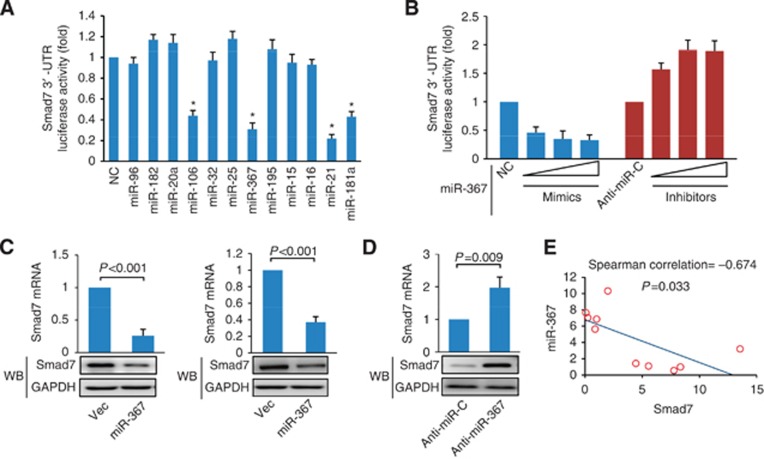
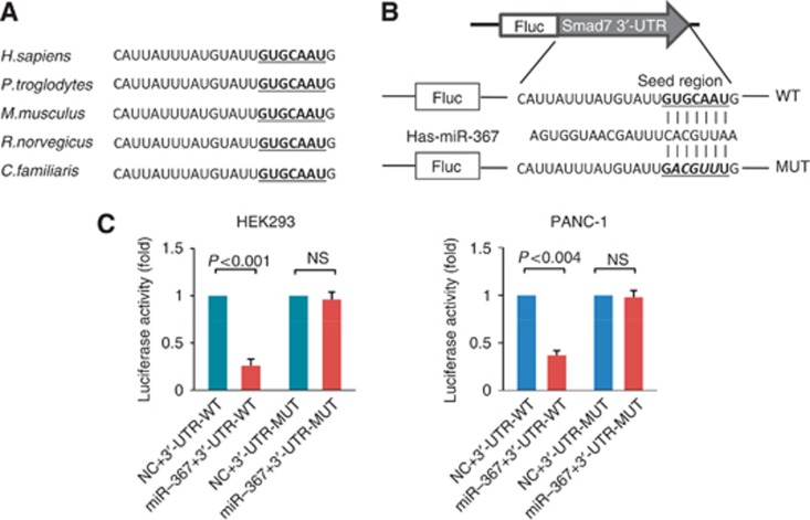
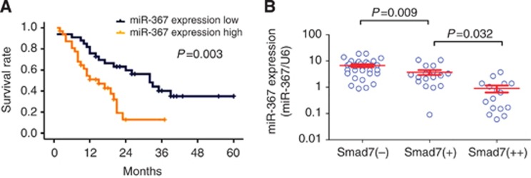
Methods: Bioinformatic prediction programmes and luciferase reporter assay were used to identify microRNAs regulating Smad7. The association between miR-367 expression and the overall survival of PDAC patients was evaluated by Kaplan-Meier analysis. The effects of miR-367 and Smad7 on the invasion and metastasis of pancreatic cancer cells were investigated both in vitro and in vivo.

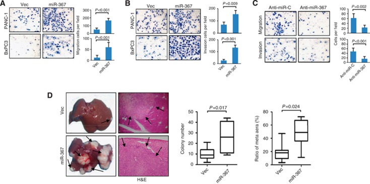
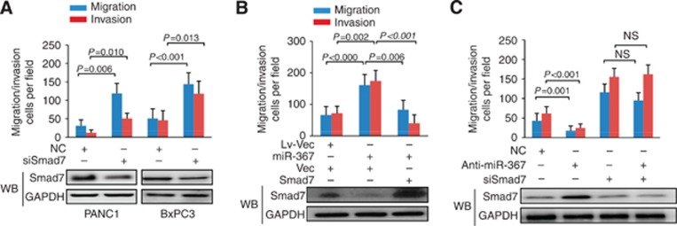
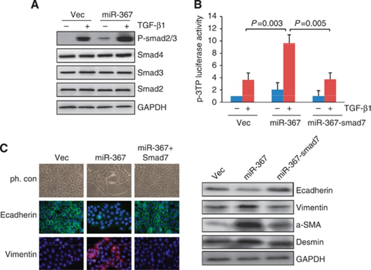
Results: We found that miR-367 downregulated Smad7 expression by directly targeting its 3'-UTR in human pancreatic cancer cells. High level of miR-367 expression correlated with poor prognosis of PDAC patients. Functional studies showed that miR-367 promoted pancreatic cancer invasion in vitro and metastasis in vivo through downregulating Smad7. In addition, we showed that miR-367 promoted epithelial-to-mesenchymal transition by increasing transforming growth factor-β (TGF-β)-induced transcriptional activity.

Conclusions: The present study identified and characterised a signalling pathway, the miR-367/Smad7-TGF-β pathway, which is involved in the invasion and metastasis of pancreatic cancer cells. Our results suggest that miR-367 may be a promising therapeutic target for the treatment of human pancreatic cancer.


Figures

Figure 1
Figure 1
Identification of miR-367 as a negative regulator of Smad7 in human pancreatic cancer cells. (A) HEK293 cells were co-transfected with the negative control (NC) or miRNA mimics and the luciferase reporter construct containing the wild-type Smad7 3′-UTR. The results were normalised to the luciferase activity detected in hek293 cells transfected with the NC, and the luciferase activity of the NC was set to 1. NS, not significant. Statistical significance was evaluated using ANOVA test. *P<0.05. (B) Mimics (20, 40, and 60 nM) or inhibitors (60, 80, and 100 nM) of miR-367 were co-transfected into PANC-1 cells with the luciferase reporter construct containing the wild-type Smad7 3′-UTR. A scrambled mimic (60 nM, NC) or inhibitor (100 nM, Anti-miR-C) was used as a control. (C) Pancreatic cancer cells were infected with control or miR-367-expressing lentiviral vector. The expression levels of Smad7 mRNA and protein were evaluated using qRT–PCR and western blot analysis, respectively. (D) Pancreatic cancer cells were transiently transfected with anti-miR-367 inhibitor or anti-miR-C (NC). Twenty-four hours later, the expression levels of Smad7 mRNA and protein were evaluated using qRT–PCR and western blot analysis, respectively. Student's t-test was used to analyse the statistical significance of differences between groups. (E) The correlation between miR-367 and Smad7 mRNA expressions in pancreatic cancer biopsies obtained from pancreatic cancer patients (n=10) was evaluated using Spearman's correlation analysis.
Figure 2
Figure 2
miR-367 downregulated Smad7 expression by directly targeting its 3′-UTR. (A) The sequences on Smad7 3′-UTR for potential binding with miR-367 were aligned among human (H. sapiens), chimpanzee (P. troglodytes), mouse (M. musculus), rat (R. norvegicus), and dog (C. familiaris). Seed sequences were highlighted and underlined. (B) A human Smad7 3′-UTR fragment containing the wild-type or mutant miR-367-binding sequence was cloned downstream to the luciferase reporter gene. (C) HEK293 and PANC-1 cells were co-transfected with the negative control (NC) or miR-367 mimics and the luciferase reporter construct containing the wild-type or mutant Smad7 3′-UTR. For each experiment, the results were normalised to the luciferase activity detected in the cells transfected with the control vector. The luciferase activity of the control vector was set to 1. NS, not significant. Statistical significance was evaluated using Student's t-test.
Figure 3
Figure 3
miR-367 expression correlated with the prognosis of PDAC. (A) Kaplan–Meier survival analysis of PDAC patients grouped by the expression levels of miR-367. The expression levels of mature miR-367 were evaluated by TaqMan real-time qPCR, and the median value of all 65 cases was defined as the cutoff value classified 65 tumour cases into low expression (n=33) and high expression of miR-367 (n=32). (B) The correlation between the expressions of Smad7 and miR-367 was assessed in 65 PDAC patients. Smad7 protein expression was evaluated by immunohistochemistry (Wang et al, 2009b). Statistical analysis of the difference in miR-367 levels between Smad7(−) and Smad7(+) samples was conducted using ANOVA test.
Figure 4
Figure 4
miR-367 promoted the invasion and metastasis of pancreatic cancer cells. (A) Migration and (B) invasion assays were performed using control lentiviral vector-infected or miR-367 vector-infected PANC-1 and BxPC3 cells. The number of cells that passed through the membrane was counted in 10 fields under the × 20 objective lens, original magnification ( × 200). The results obtained from three independent experiments were presented as the mean±s.d. of values. (C) Migration and invasion assays were performed using negative control (anti-miR-C) or anti-miR-367 inhibitor-transfected PANC-1 cells. The results obtained from three independent experiments were presented as the mean±s.d. of values. (D) Impact of miR-367 on tumour metastasis in vivo. Eight weeks after spleen injection of control vector-infected or miR-367 vector-infected cells, the mice were killed, and the livers were dissected. Representative livers from NOD/SCID mice were shown. H&E staining was performed on sections of metastatic tumours and normal liver tissues. Arrows represent the metastatic nodules, original magnification ( × 200). Tumour metastases were quantified by counting the number of metastatic colonies in one histological section of the midportion of liver specimen from mice. The ratio of the metastatic area to the total area was determined in histological sections from the midportion of each liver (7 per group). Student's t-test was used to analyse the statistical significance of the differences between groups.
Figure 5
Figure 5
Smad7 mediated the invasion-promotion function of miR-367 in pancreatic cancer cells. (A) Migration and invasion assays of pancreatic cancer cells transfected with Smad7 siRNA. The expression level of Smad7 protein 24 h after transfection was evaluated by western blot analysis. The number of cells that passed through the membrane was counted in 10 fields under the × 20 objective lens, original magnification ( × 200). The results obtained from three independent experiments were presented as the mean±s.d. of values. (B) Migration and invasion assays of control vector-expressing and miR-367-expressing PANC1 cells transfected with a control vector or a Smad7-expressing vector that contained the entire coding sequence of Smad7 but lacked the 3′-UTR. The expression level of Smad7 protein was evaluated by western blot analysis. The number of cells that passed through the membrane was counted in 10 fields under the × 20 objective lens, original magnification ( × 200). The results obtained from three independent experiments were presented as the mean±s.d. of values. (C) Migration and invasion assays of PANC1 cells co-transfected with anti-miR-367 and siSmad7. Smad7 protein levels were evaluated by western blot analysis. The number of cells that invaded through the membrane was counted in 10 fields under the × 20 objective lens., original magnification, × 200. The results were presented as the mean±s.d. of values obtained from three independent experiments.
Figure 6
Figure 6
miR-367 promoted EMT by increasing TGF-β-induced transcriptional activity. (A) Control lentiviral vector-infected or miR-367 vector-infected PANC-1 cells were uninduced or induced by TGF-β1 (200 pM) for 1 h. Whole-cell extracts were analysed by western blot analysis using antibodies against Smad2, Smad3, Smad4, and phosphorylated Smad2/3. GAPDH was used as a loading control. (B) Control vector-expressing and miR-367-expressing cells were co-transfected with a Smad7-expressing vector that contained the entire coding sequence of Smad7 but lacked the 3′-UTR and a TGF-β responsive luciferase reporter, p3TP. TGF-β-induced luciferase activity was measured. The results obtained from three independent experiments were presented as the mean±s.d. of values. (C) BxPC3 cells were stained to detect E-cadherin and vimentin (green). Cell nuclei were counterstained with DAPI, original magnification ( × 200). The morphology of cells was shown by phase-contrast images (ph.con.). Total protein was extracted, and the expressions of E-cadherin, vimentin, a-SMA, and desmin were evaluated by western blot analysis. GAPDH was used as a loading control.

References

    1. Akhurst RJ, Derynck R. TGF-beta signaling in cancer—a double-edged sword. Trends Cell Biol. 2001;11:S44–S51. - PubMed
    1. Azuma H, Ehata S, Miyazaki H, Watabe T, Maruyama O, Imamura T, Sakamoto T, Kiyama S, Kiyama Y, Ubai T, Inamoto T, Takahara S, Itoh Y, Otsuki Y, Katsuoka Y, Miyazono K, Horie S. Effect of Smad7 expression on metastasis of mouse mammary carcinoma JygMC(A) cells. J Natl Cancer Inst. 2005;97:1734–1746. - PubMed
    1. Bai S, Cao X. A nuclear antagonistic mechanism of inhibitory Smads in transforming growth factor-beta signaling. J Biol Chem. 2002;277:4176–4182. - PubMed
    1. Boulay JL, Mild G, Lowy A, Reuter J, Lagrange M, Terracciano L, Laffer U, Herrmann R, Rochlitz C. SMAD7 is a prognostic marker in patients with colorectal cancer. Int J Cancer. 2003;104:446–449. - PubMed
    1. Cerutti JM, Ebina KN, Matsuo SE, Martins L, Maciel RM, Kimura ET. Expression of Smad4 and Smad7 in human thyroid follicular carcinoma cell lines. J Endocrinol Invest. 2003;26:516–521. - PubMed

Publication types

MeSH terms