. 2015 Apr 13;10(4):e0123152.
doi: 10.1371/journal.pone.0123152. eCollection 2015.

TLR-3 is present in human adipocytes, but its signalling is not required for obesity-induced inflammation in adipose tissue in vivo

Affiliations

TLR-3 is present in human adipocytes, but its signalling is not required for obesity-induced inflammation in adipose tissue in vivo

Dov B Ballak et al. PLoS One. .

Abstract

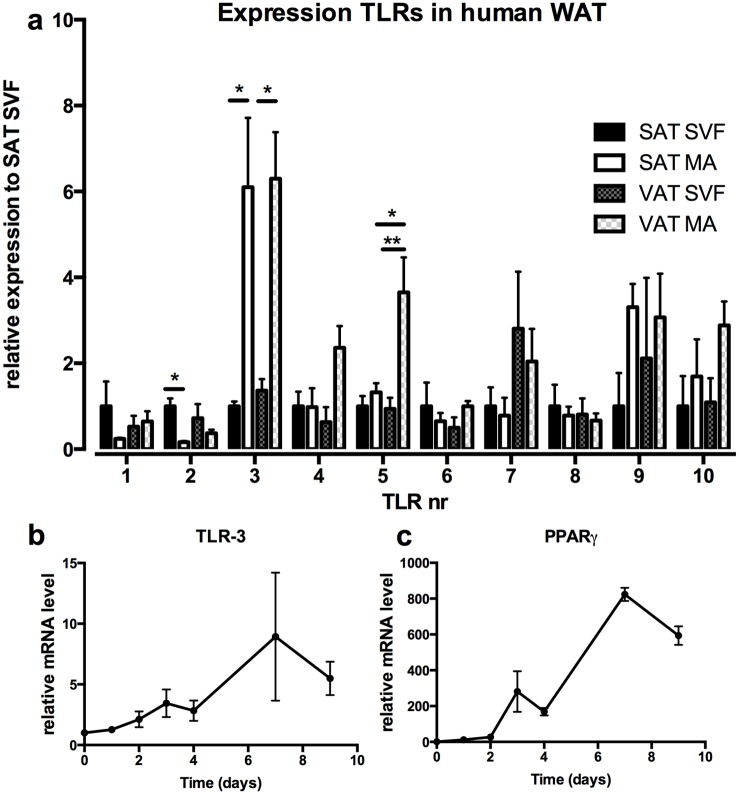
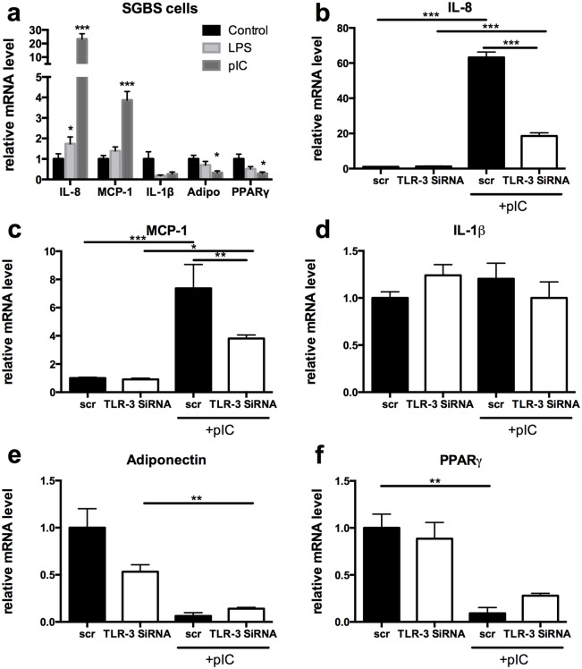
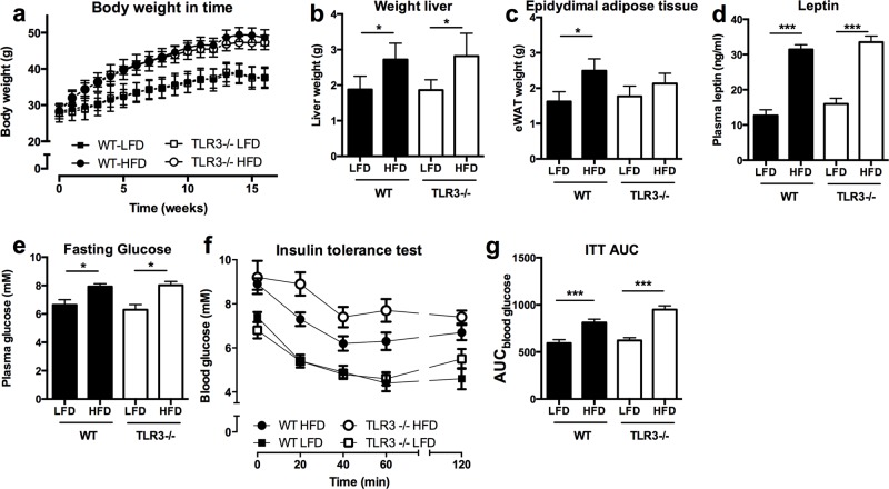
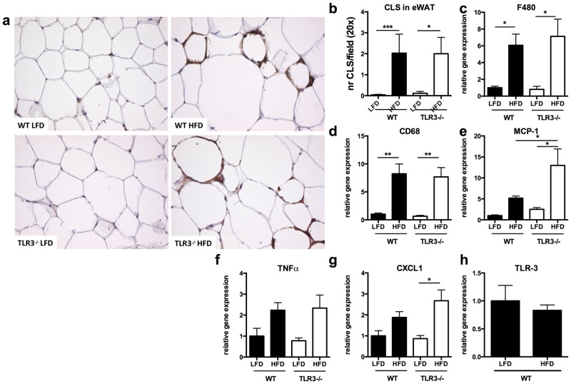
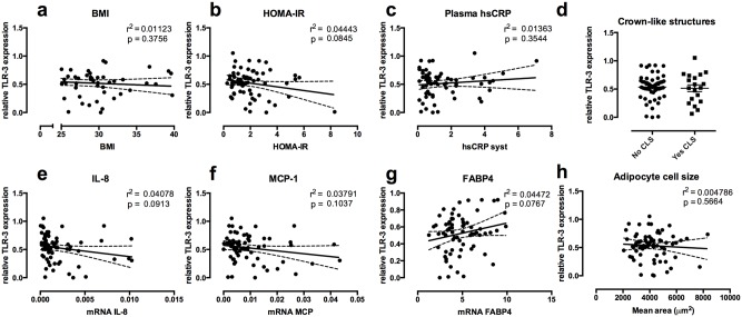
Innate immunity plays a pivotal role in obesity-induced low-grade inflammation originating from adipose tissue. Key receptors of the innate immune system including Toll-like receptors-2 and -4 (TLRs) are triggered by nutrient excess to promote inflammation. The role of other TLRs in this process is largely unknown. In addition to double-stranded viral mRNA, TLR-3 can also recognize mRNA from dying endogenous cells, a process that is frequently observed within obese adipose tissue. Here, we identified profound expression of TLR-3 in adipocytes and investigated its role during diet-induced obesity. Human adipose tissue biopsies (n=80) and an adipocyte cell-line were used to study TLR-3 expression and function. TLR-3-/- and WT animals were exposed to a high-fat diet (HFD) for 16 weeks to induce obesity. Expression of TLR-3 was significantly higher in human adipocytes compared to the non-adipocyte cells part of the adipose tissue. In vitro, TLR-3 expression was induced during differentiation of adipocytes and stimulation of the receptor led to elevated expression of pro-inflammatory cytokines. In vivo, TLR-3 deficiency did not significantly influence HFD-induced obesity, insulin sensitivity or inflammation. In humans, TLR-3 expression in adipose tissue did not correlate with BMI or insulin sensitivity (HOMA-IR). Together, our results demonstrate that TLR-3 is highly expressed in adipocytes and functionally active. However, TLR-3 appears to play a redundant role in obesity-induced inflammation and insulin resistance.


Conflict of interest statement

Competing Interests: The authors have declared that no competing interests exist.

Figures

Fig 1
Fig 1. TLR-3 is predominantly expressed in adipocytes.
(a) Biopsies from visceral- (VAT) and subcutaneous adipose tissue (SAT) were obtained from 4 healthy subjects and TLR expression was determined in stromal vascular fraction (SVF) and mature adipocytes (MA). mRNA levels of (b) TLR-3 and (c) PPAR-γ were measured during differentiation of human SGBS adipocytes. * p<0.05, ** p<0.01. Data are shown as means ± SEM.
Fig 2
Fig 2. TLR-3 is functionally active in adipocytes.
(a) Differentiated SGBS adipocytes were stimulated with either a TLR-3 (poly:IC 12.5μg/ml) or TLR-4 (LPS 50ng/ml) agonist. mRNA levels were measured for IL-8, MCP-1, IL-1 β, adiponectin and PPAR-γ. (b-f) SGBS adipocytes were treated with SiRNA against TLR-3 or scr SiRNA and stimulated with poly:IC. mRNA levels of (b) IL-8, (c) MCP-1, (d) IL-1β, (e) adiponectin, (f) PPAR- γ were subsequently measured. * p<0.05, ** p<0.01, *** p<0.001. Data are shown as means ± SEM.
Fig 3
Fig 3. TLR-3 deficiency does not protect mice against metabolic abnormalities.
Wild-type (WT) and TLR-3-/- mice were subjected to 16 weeks of low fat diet (LFD) or high fat diet (HFD). (a) development of the bodyweight, (b) liver weight, (c) epididymal adipose tissue weight, (d) plasma leptin levels, (e) fasting glucose levels, (f) insulin tolerance test (ITT), (g) area under the curve for ITT. * p<0.05, *** p<0.001. Number of mice per group: WT-LFD n = 10; WT-HFD n = 10; TLR-3-/-LFD n = 7; TLR-3-/-HFD n = 9. Data are shown as means ± SEM.
Fig 4
Fig 4. TLR-3 deficiency does not ameliorate adipose tissue inflammation.
After 16 weeks of low fat diet (LFD) or high fat diet (HFD) intervention, adipose tissue of wild-type (WT) and TLR-3-/- mice was investigated for inflammatory parameters. (a) Adipose tissue of mice stained for F4/80, magnification 20x, (b) number of crown-like structures in adipose tissue. Inflammatory markers were measured (c) F480, (d) CD68, (e) MCP-1, (f) TNFα, (g) CXCL1. (h) mRNA levels of TLR-3 in WT mice fed a LFD or HFD for 16 weeks. * p<0.05, ** p<0.01. Number of mice per group: WT-LFD n = 10; WT-HFD n = 10; TLR-3-/-LFD n = 7; TLR-3-/-HFD n = 9. Data are shown as means ± SEM.
Fig 5
Fig 5. TLR-3 in human adipose tissue.
Subcutaneous adipose tissue samples of 80 healthy individuals were obtained. TLR-3 mRNA levels were associated with (a) BMI, (b) HOMA-IR, (c) plasma CRP-levels, (d) number of crown-like structures in adipose tissue. Association of MAP3K8 mRNA expression in human subcutaneous adipose tissue with mRNA expression of (e) IL-8, (f) MCP-1, (g) FABP4 and (h) adipocytes cell size. * p<0.05. Data are shown as means ± SEM. HOMA-IR = Homeostatic Model Assessment for insulin resistance.

References

    1. WHO. WHO fact files: ten facts on obesity. 2010. Geneva: WHO.
    1. Donath MY, Shoelson SE. Type 2 diabetes as an inflammatory disease. Nat Rev Immunol. 2011; 11: 98–107. 10.1038/nri2925 - DOI - PubMed
    1. Weisberg SP, McCann D, Desai M, Rosenbaum M, Leibel RL, Ferrante AW Jr. Obesity is associated with macrophage accumulation in adipose tissue. J Clin Invest. 2003; 112: 1796–1808. - PMC - PubMed
    1. Xu H, Barnes GT, Yang Q, Tan G, Yang D, Chou CJ, et al. Chronic inflammation in fat plays a crucial role in the development of obesity-related insulin resistance. J Clin Invest. 2003; 112: 1821–1830. - PMC - PubMed
    1. Duffaut C, Galitzky J, Lafontan M, Bouloumie A. Unexpected trafficking of immune cells within the adipose tissue during the onset of obesity. Biochem Biophys Res Commun. 2009; 384: 482–485. 10.1016/j.bbrc.2009.05.002 - DOI - PubMed

Publication types