. 2015 Jun;95(6):684-96.
doi: 10.1038/labinvest.2015.51. Epub 2015 Apr 13.

Development and characterization of human-induced pluripotent stem cell-derived cholangiocytes

Affiliations

Development and characterization of human-induced pluripotent stem cell-derived cholangiocytes

Thiago M De Assuncao et al. Lab Invest. 2015 Jun.

Erratum in

Abstract

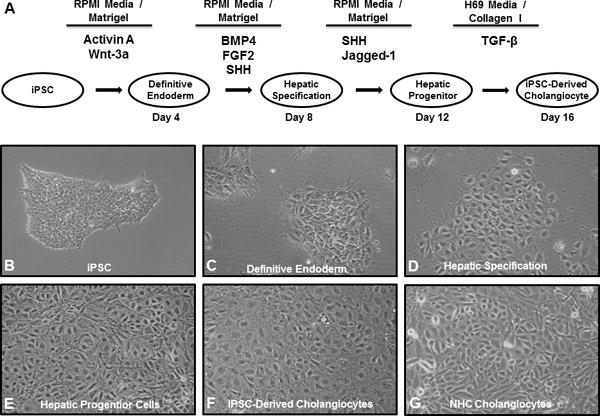
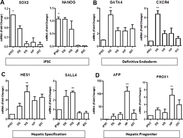
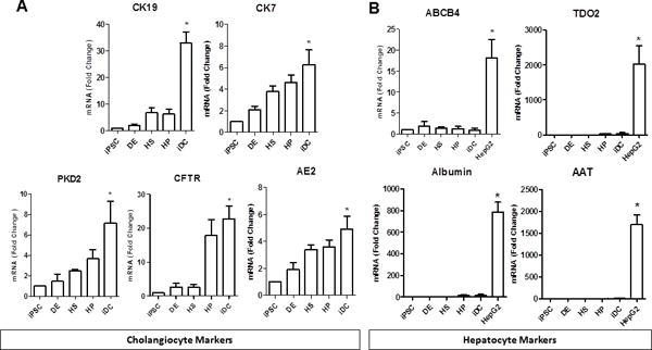
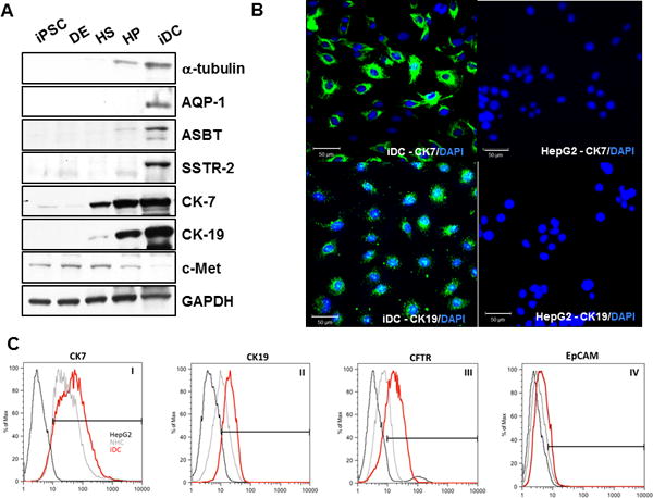
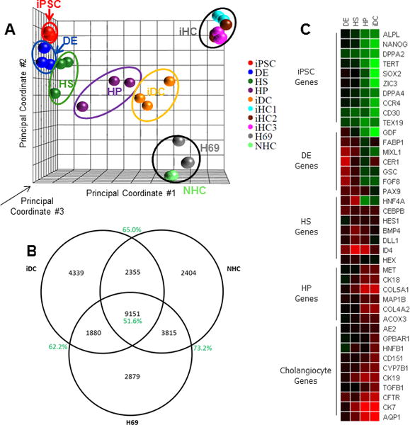
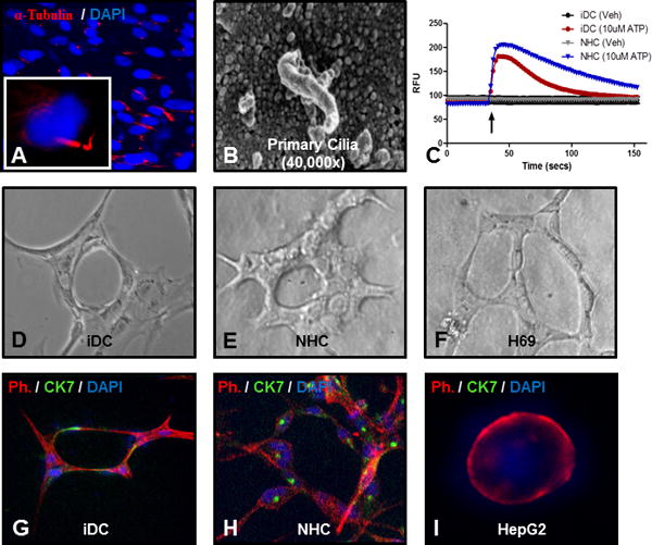
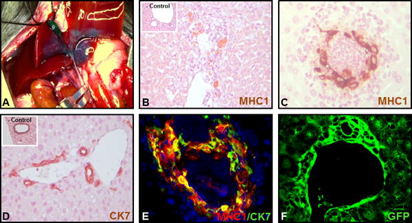
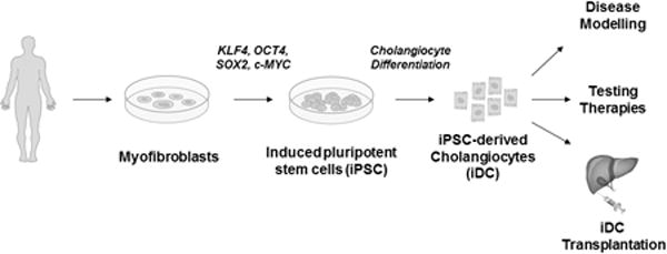
Cholangiocytes are the target of a heterogeneous group of liver diseases known as the cholangiopathies. An evolving understanding of the mechanisms driving biliary development provides the theoretical underpinnings for rational development of induced pluripotent stem cell (iPSC)-derived cholangiocytes (iDCs). Therefore, the aims of this study were to develop an approach to generate iDCs and to fully characterize the cells in vitro and in vivo. Human iPSC lines were generated by forced expression of the Yamanaka pluripotency factors. We then pursued a stepwise differentiation strategy toward iDCs, using precise temporal exposure to key biliary morphogens, and we characterized the cells, using a variety of morphologic, molecular, cell biologic, functional, and in vivo approaches. Morphology shows a stepwise phenotypic change toward an epithelial monolayer. Molecular analysis during differentiation shows appropriate enrichment in markers of iPSC, definitive endoderm, hepatic specification, hepatic progenitors, and ultimately cholangiocytes. Immunostaining, western blotting, and flow cytometry demonstrate enrichment of multiple functionally relevant biliary proteins. RNA sequencing reveals that the transcriptome moves progressively toward that of human cholangiocytes. iDCs generate intracellular calcium signaling in response to ATP, form intact primary cilia, and self-assemble into duct-like structures in three-dimensional culture. In vivo, the cells engraft within mouse liver, following retrograde intrabiliary infusion. In summary, we have developed a novel approach to generate mature cholangiocytes from iPSCs. In addition to providing a model of biliary differentiation, iDCs represent a platform for in vitro disease modeling, pharmacologic testing, and individualized, cell-based, regenerative therapies for the cholangiopathies.


Conflict of interest statement

Disclosure/Duality of Interest

The authors have nothing to disclose.

Figures

Figure 1
Figure 1. Protocol for generation of iDC from iPSC and morphologic analysis
(A) The protocol proceeds through phases of DE, HS, and HP and incorporates defined culture media, precise extracellular components, and temporal exposure to key biliary morphogens. Light microscopy (10X) shows phenotypic changes at each stage of differentiation with the transformation of (B) the stem cell phenotype towards (C–F) an epithelial monolayer, similar to the (G) NHC cholangiocyte cell line.
Figure 2
Figure 2. Molecular analysis
Real-time PCR showing expression of (A) pluripotency markers (Sox2 and NANOG) in iPSCs, followed by (B) expression of definitive endoderm markers (GATA4 and CXCR4), (C) hepatic specification markers (Hes1 and Sall4), and (D) hepatic progenitor markers (AFP and PROX1). *p≤0.05 compared to iDC, **p≤0.05 compared to iPSC. All data is normalized to GAPDH and iPSC expression is set to 1.0.
Figure 3
Figure 3. Cholangiocyte gene expression
Real-time PCR shows that (A) iDCs are enriched in the cholangiocyte markers CK19, CK7, CFTR, PKD2, and AE2. iDCs lack expression of (B) hepatocyte markers, ABCB4, albumin, α-1-antitrypsin (A1AT), and tryptophan 2,3-dioxygenase (TDO2), *p≤0.05 compared to iPSC. All data is normalized to GAPDH and iPSC expression is set to 1.0.
Figure 4
Figure 4. Expression of cholangiocyte proteins
(A) Western blotting shows enrichment of multiple, functionally relevant biliary proteins in iDCs including acetylated α-tubulin, AQP-1, ASBT, SSTR-2, CK-7, and CK-19, while c-Met was not significantly detected. (B) iDCs were stained for CK7 and CK19 by immunofluorescence (left panels). HepG2 hepatocytes showed no staining for either marker (right panels). (C) Flow cytometry analyses of choalngiocyte markers, including CK7, CK19 and CFTR (panels I, II and III, respectively) and hepatic endoderm marker EpCAM (panel IV). Scale bars = 50 μm
Figure 5
Figure 5. RNA sequencing
Sequencing of the entire transcriptome was performed at each phase of differentiation and compared to the transpcriptome of cholangiocyte cell lines and isolated human choloangiocytes. (A) Principal component analysis provided a 3-dimensional graphical representation of the gene expression clustering between groups based on three mathematically-defined principal components. Triplicate samples are contained within the ovals. We see a smooth transition from iPSC (red) to iDC (gold) with a trajectory toward the H69 (grey), NHC (green), and iHC (light blue, brown, and purple) cholangiocytes. (B) Differential expression analysis, performed relative to iPSC, displayed as a Venn diagram, confirms similarly regulated genes between iDCs, H69 cells, and NHCs. (C) A heatmap showing fold-change relative to iPSC demonstrates appropriate enrichment in developmental genes as differentiation progresses.
Figure 6
Figure 6. Functional characteristics of iDCs
iDCs were able to form primary cilia as shown by (A) immunofluorescence for acetylated α-tubulin (red) and DAPI (blue) or by (B) scanning electron microscopy. (C) iDCs activate intracellular calcium signaling in response to ATP. (D) iDCs form round duct-like structures with a luminal space when cultured for 7 days in a 3D type I collagen / Matrigel culture system, similar to (E) NMC cells (F) H69 cells. (G) Co-staining with phalloidin (red), CK7 (green) and DAPI (blue) confirms CK7 positivity in the duct-like structures, similar to (H) NHC cells, but different from (I) HepG2 hepatocytes.
Figure 7
Figure 7. Retrograde intrabiliary infusion
(A) Retrograde infusion of iDCs was accomplished by microsurgical cannulation of the cystic duct with temporary occlusion of the common bile duct. Engraftment was analyzed by (B–C) staining for human class 1 MHC (inset shows a cell-free control injection) or (D) CK7 (inset shows a cell-free control injection), by (E) dual label immunofluorescence for MHC1 (red) and CK7 (green), and if cells are pre-labelled with GFP, by (F) confocal microscopy for GFP.
Figure 8
Figure 8. Development and potential uses for iDC
The flowchart outlines the development of iPSC from myofibroblasts, differentiation to iDCs, and potential downstream applications for the cells.

References

    1. Lazaridis KN, Strazzabosco M, Larusso NF. The cholangiopathies: disorders of biliary epithelia. Gastroenterology. 2004;127(5):1565–1577. - PubMed
    1. O’Hara SP, Tabibian JH, Splinter PL, LaRusso NF. The dynamic biliary epithelia: molecules, pathways, and disease. J Hepatol. 2013;58(3):575–582. - PMC - PubMed
    1. Kochhar G, Parungao JM, Hanouneh IA, Parsi MA. Biliary complications following liver transplantation. World J Gastroenterol. 2013;19(19):2841–2846. - PMC - PubMed
    1. Mourad MM, Algarni A, Liossis C, Bramhall SR. Aetiology and risk factors of ischaemic cholangiopathy after liver transplantation. World J Gastroenterol. 2014;20(20):6159–6169. - PMC - PubMed
    1. Williams MJ, Clouston AD, Forbes SJ. Links between hepatic fibrosis, ductular reaction, and progenitor cell expansion. Gastroenterology. 2014;146(2):349–356. - PubMed

Publication types

MeSH terms