Review
. 2015 Sep;26(9):2074-80.
doi: 10.1681/ASN.2014121274. Epub 2015 Apr 13.

Paradigm Shifts in Atherosclerotic Renovascular Disease: Where Are We Now?

Affiliations
Review

Paradigm Shifts in Atherosclerotic Renovascular Disease: Where Are We Now?

Stephen C Textor et al. J Am Soc Nephrol. 2015 Sep.

Abstract

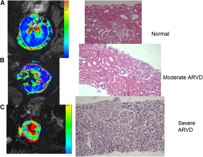
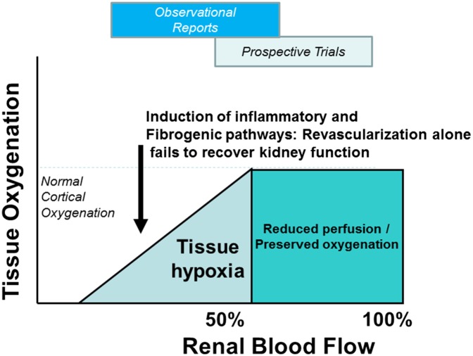
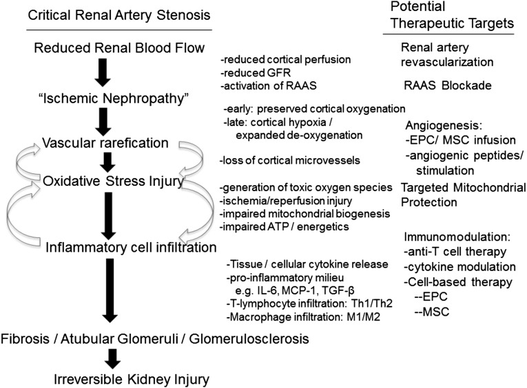
Results of recent clinical trials and experimental studies indicate that whereas atherosclerotic renovascular disease can accelerate both systemic hypertension and tissue injury in the poststenotic kidney, restoring vessel patency alone is insufficient to recover kidney function for most subjects. Kidney injury in atherosclerotic renovascular disease reflects complex interactions among vascular rarefication, oxidative stress injury, and recruitment of inflammatory cellular elements that ultimately produce fibrosis. Classic paradigms for simply restoring blood flow are shifting to implementation of therapy targeting mitochondria and cell-based functions to allow regeneration of vascular, glomerular, and tubular structures sufficient to recover, or at least stabilize, renal function. These developments offer exciting possibilities of repair and regeneration of kidney tissue that may limit progressive CKD in atherosclerotic renovascular disease and may apply to other conditions in which inflammatory injury is a major common pathway.

Keywords: macrophages; mesangial cells; oxidative stress; renal artery stenosis.


Figures

Figure 1.
Figure 1.
Cortical hypoxia and inflammation develop in severe ARVD. Parametric axial image maps of deoxyhemoglobin determined by BOLD MR (left panels) and transjugular or needle biopsy samples from (A) a normal subject (kidney donor implantation biopsy), (B) an individual with moderate ARVD (reduced blood flow and GFR, peak ultrasound velocity 350 cm/s), and (C) severe ARVD with loss of cortical volume and function and peak ultrasound velocity >450 cm/s. Panel (B) shows that despite reduced blood flow, tissue oxygenation in the cortex and medullary segments are remarkably preserved, consistent with near normal histologic appearance and stable renal function during antihypertensive drug therapy. Panel (C) demonstrates higher levels of cortical deoxyhemoglobin (green) associated with more severe ARVD, along with a larger fraction of the medullary segments being overtly hypoxic with elevated (yellow/red) deoxyhemoglobin. These findings were associated with histologic changes of interstitial inflammatory cell infiltrates (see additional information in the text) and tubular degeneration.
Figure 2.
Figure 2.
Clinical results depend on the degree of blood flow reduction tissue hypoxia and the level of inflammatory and fibrotic injury. Schematic summary of the working paradigms related to ARVD. As a filtering organ, the kidney can adapt to moderate reductions of blood flow with no loss of tissue oxygenation. When one exceeds the lower limits of adaptation, eventual tissue hypoxia develops associated with activation of multiple injury pathways that may not be affected by simply restoring vessel patency and blood flow. Substantial differences between observational reports of ARVD and results from prospective trials likely reflect a degree of selection bias favoring moderate disease on the basis of vascular anatomy and hemodynamics alone (see additional information in the text).
Figure 3.
Figure 3.
Injury pathways and targets in ARVD. Working diagram highlighting recent experimental studies delineating specific pathways of oxidative stress injury and inflammatory injury pathways in the poststenotic kidney. The right panel identifies specific therapeutic targets that may alleviate these injury pathways, over and above simply restoring blood flow (see additional information in the text). Many of these pathways likely overlap in many forms of kidney injury. EPC, endothelial progenitor cells; MCP, monocyte chemoattractant protein.

References

    1. Textor SC: Atherosclerotic renal artery stenosis: Overtreated but underrated? J Am Soc Nephrol 19: 656–659, 2008 - PubMed
    1. Murphy TP, Soares G, Kim M: Increase in utilization of percutaneous renal artery interventions by medicare beneficiaries, 1996-2000. AJR Am J Roentgenol 183: 561–568, 2004 - PubMed
    1. Balk E, Raman G, Chung M, Ip S, Tatsioni A, Alonso A, Chew P, Gilbert SJ, Lau J: Effectiveness of management strategies for renal artery stenosis: A systematic review. Ann Intern Med 145: 901–912, 2006 - PubMed
    1. Herrmann SM, Saad A, Textor SC: Management of atherosclerotic renovascular disease after Cardiovascular Outcomes in Renal Atherosclerotic Lesions (CORAL). Nephrol Dial Transplant 30: 366–375, 2015 - PMC - PubMed
    1. Ritchie J, Green D, Chrysochou C, Chalmers N, Foley RN, Kalra PA: High-risk clinical presentations in atherosclerotic renovascular disease: Prognosis and response to renal artery revascularization. Am J Kidney Dis 63: 186–197, 2014 - PubMed

Publication types