. 2015 Sep;38(5):905-14.
doi: 10.1007/s10545-015-9836-6. Epub 2015 Apr 14.

Clinical, morphological, biochemical, imaging and outcome parameters in 21 individuals with mitochondrial maintenance defect related to FBXL4 mutations

Affiliations

Clinical, morphological, biochemical, imaging and outcome parameters in 21 individuals with mitochondrial maintenance defect related to FBXL4 mutations

Martina Huemer et al. J Inherit Metab Dis. 2015 Sep.

Abstract

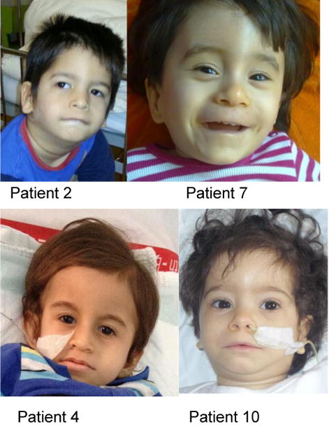
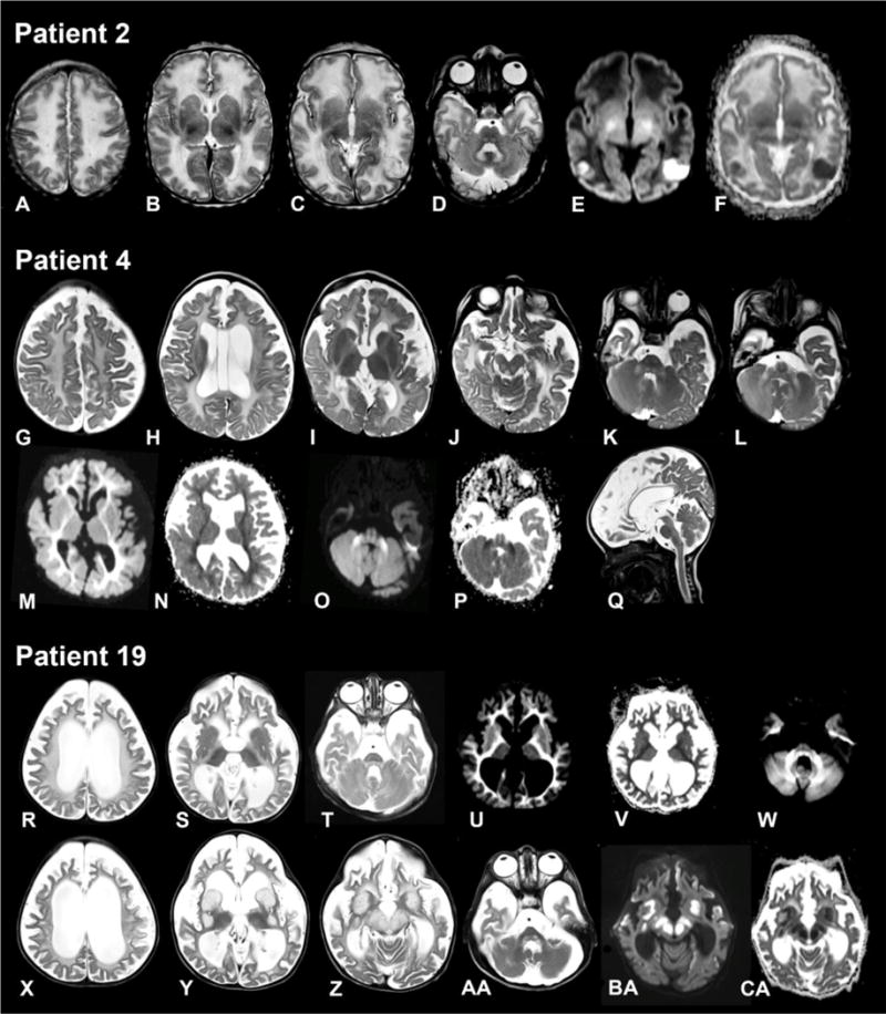
FBXL4 deficiency is a recently described disorder of mitochondrial maintenance associated with a loss of mitochondrial DNA in cells. To date, the genetic diagnosis of FBXL4 deficiency has been established in 28 individuals. This paper retrospectively reviews proxy-reported clinical and biochemical findings and evaluates brain imaging, morphological and genetic data in 21 of those patients. Neonatal/early-onset severe lactic acidosis, muscular hypotonia, feeding problems and failure to thrive is the characteristic pattern at first presentation. Facial dysmorphic features are present in 67% of cases. Seven children died (mean age 37 months); 11 children were alive (mean age at follow-up 46 months), three children were lost to follow-up. All survivors developed severe psychomotor retardation. Brain imaging was non-specific in neonates but a later-onset, rapidly progressive brain atrophy was noted. Elevated blood lactate and metabolic acidosis were observed in all individuals; creatine kinase was elevated in 45% of measurements. Diagnostic workup in patient tissues and cells revealed a severe combined respiratory chain defect with a general decrease of enzymes associated with mitochondrial energy metabolism and a relative depletion of mitochondrial DNA content. Mutations were detected throughout the FBXL4 gene albeit with no clear delineation of a genotype-phenotype correlation. Treatment with "mitochondrial medications" did not prove effective. In conclusion, a clinical pattern of early-onset encephalopathy, persistent lactic acidosis, profound muscular hypotonia and typical facial dysmorphism should prompt initiation of molecular genetic analysis of FBXL4. Establishment of the diagnosis permits genetic counselling, prevents patients undergoing unhelpful diagnostic procedures and allows for accurate prognosis.


Conflict of interest statement

Compliance with ethics guidelines Conflict of interest None.

Figures

Fig. 1
Fig. 1
Main clinical features (columns) and number of available ratings for each parameter (lines) at first presentation and ever during the course in 21 patients with FBXL4 mutations
Fig. 2
Fig. 2. Patients with FBXL4 deficiency share typical facial characteristics
Patient 2: Prominent, narrow forehead with intertemporal narrowing, thick, medial diffuse eyebrows with slight synophris, bilateral epicanthus, periorbital fullness, broad nasal root and broad nasal ridge, fullness of paranasal tissue, flat malar bones, long philtrum, large, low set, protruding ears with overfolded helices. Patient 7: Prominent forehead, thick, diffuse eyebrows, short, upslanting palpebral fissures, broad nasal root, depressed nasal bridge, wide nasal base, smooth philtrum, thin vermillion of the upper lip, low set ears. Patient 4: Broad, prominent forehead, thick eyebrows, short palpebral fissures, broad nasal root, wide nasal base, low set ears. Patient 10: Prominent forehead, medial diffuse eyebrows with slight synophris, short, upslanting palpebral fissures, broad nasal root, depressed nasal bridge, wide nasal base, smooth philtrum, thin vermillion of the upper lip, low set, protruding ears.
Fig. 3
Fig. 3. Magnetic resonance imaging of the brain from patient 2 (4 days), patient 4 (9 months) and patient 19 (3 years)
Patient 2 (age 4 days). Axial T2-weighted images (A–D) show swollen and slightly hyperintense aspect of supratentorial white matter. There are several defects in the basal ganglia (B). There are two small areas at the parietooccipital border on both sides where the cortex is less well defined (C) with corresponding impaired diffusion (E) and decreased apparent diffusion coefficient (ADC) (F). Signal of the dorsal tegmental tracts and hilus of the dentate nucleus is increased (D). In the supratentorial white matter, diffusivity is increased (E) with elevated ADC (F), with exception of the posterior limb of the internal capsule. Patient 4 (age 9 months). Axial T2-weighted images (G–L) demonstrate global supratentorial atrophy, a cavum vergae and a left paraventricular cyst (H) and a mild diffuse increase of the supratentorial white matter signal (G–I). The tectum (J), pyramidal tracts, dorsal tegmentum (K) and middle cerebellar peduncles (K, L) have an elevated signal. Diffusion is impaired with decreased ADC in the supratentorial white matter (M, N) and the middle cerebellar peduncles, dorsal tegmental tracts and pyramidal tracts (O, P). The sagittal T2-weighted image (Q) shows normal volume of the cerebellum. Patient 19 aged 3 years (R–W) and 2.5 months later (X-CA). Severe supratentorial atrophy is evident on the axial T2-weighted images (R–T), with now also thinning of the brain stem (T). White matter signal is diffusely elevated (R–T) with impaired diffusion (U, W) and a corresponding low ADC (V). 2.5 months later, basal ganglia (Y), substantia nigra and red nucleus (Z) are grossly swollen and hyperintense with corresponding impaired diffusion (BA) and ADC (CA). Also the dorsal tegmental tracts are swollen (AA).
Fig. 4
Fig. 4. FBXL4 Mutation Status and Gene Structure
(A) Gene structure of FBXL4 with localization of mutations in 28 patients. Known functional domains are colored in green and red, regions of unknown function in grey. Red and blue asterisks indicate post-translational modifications. Mutations labeled in bold origin from patients reported in this study. (B) Conservation of amino acid residues affected by mutations. Numbering and coloring correspond to NP_001265645 alignment.

References

    1. Acham-Roschitz B, Plecko B, Lindbichler F, Bittner R, Mache CJ, Sperl W, Mayr JA. A novel mutation of the RRM2B gene in an infant with early fatal encephalomyopathy, central hypomyelination, and tubulopathy. Mol Genet Metab. 2009;98:300–304. - PubMed
    1. Barbier A, Boivin A, Yoon W, et al. New reference curves for head circumference at birth, by gestational age. New reference curves for head circumference at birth, by gestational age. Pediatrics. 2013;131:1158–1167. - PubMed
    1. Berger A, Mayr JA, Meierhofer D, et al. Severe depletion of mitochondrial DNA in spinal muscular atrophy. Acta Neuropathol. 2003;105:245–251. - PubMed
    1. Blau N, Duran M, Blaskovic ME, Gibson KM, editors. Physician’s guide to the laboratory diagnosis of metabolic diseases. 2nd. Springer; Berlin: 2003.
    1. Böhm M, Pronicka E, Karcmarewicz E, et al. Retrospective, multicentric study of 180 children with cytochrome c oxidase deficiency. Pediatr Res. 2006;59:21–26. - PubMed

Publication types

MeSH terms