

Paraseptal emphysema: Prevalence and distribution on CT and association with interstitial lung abnormalities
- PMID: 25868675
- PMCID: PMC4450117
- DOI: 10.1016/j.ejrad.2015.03.010
Paraseptal emphysema: Prevalence and distribution on CT and association with interstitial lung abnormalities
Abstract
Objective: To investigate the prevalence and distribution of paraseptal emphysema on chest CT images in the Framingham Heart Study (FHS) population, and assess its impact on pulmonary function. Also pursued was the association with interstitial lung abnormalities.
Materials and methods: We assessed 2633 participants in the FHS for paraseptal emphysema on chest CT. Characteristics of the participants, including age, sex, smoking status, clinical symptoms, and results of pulmonary function tests, were compared between those with and without paraseptal emphysema. The association between paraseptal emphysema and interstitial lung abnormalities was investigated.
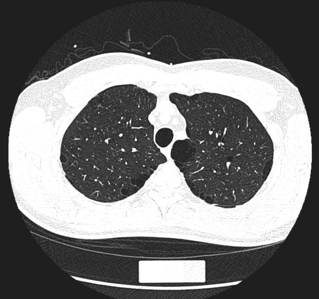
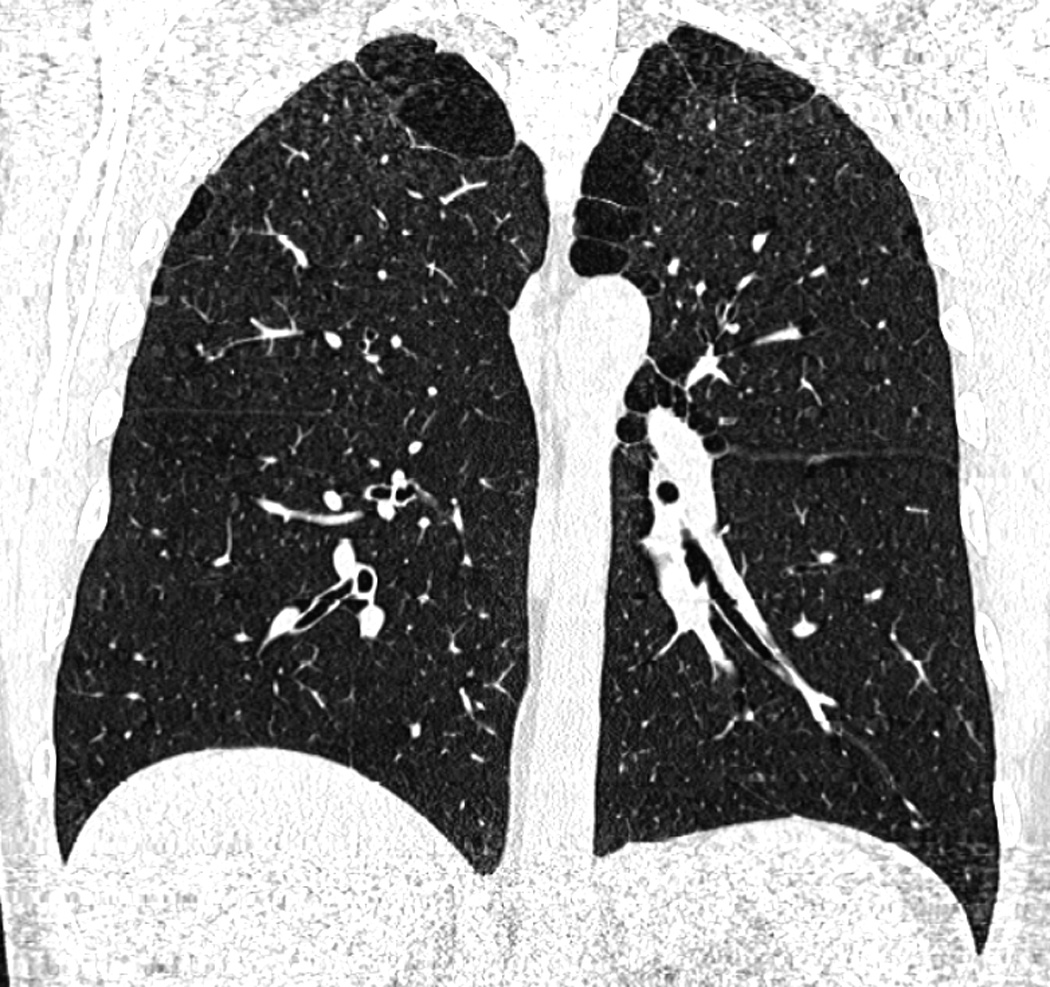
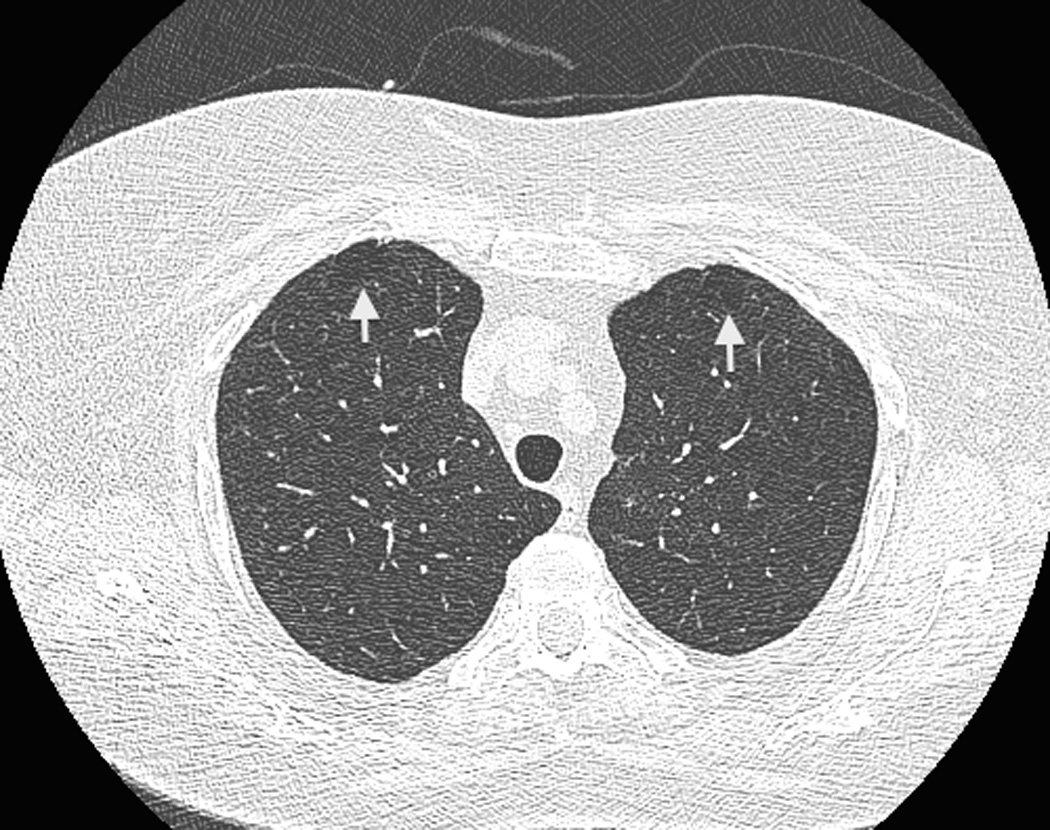
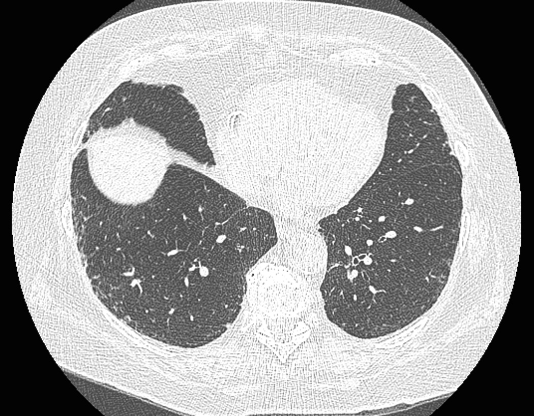
Results: Of the 2633 participants, 86 (3%) had pure paraseptal emphysema (defined as paraseptal emphysema with no other subtypes of emphysema other than paraseptal emphysema or a very few centrilobular emphysema involved) in at least one lung zone. The upper zone of the lungs was almost always involved. Compared to the participants without paraseptal emphysema, those with pure paraseptal emphysema were significantly older, and were more frequently male and smokers (mean 64 years, 71% male, mean 36 pack-years, P<0.001) and had significantly decreased FEV1/FVC% (P=0.002), and diffusion capacity of carbon monoxide (DLCO) (P=0.002). There was a significant association between pure paraseptal emphysema and interstitial lung abnormalities (P<0.001).
Conclusions: The prevalence of pure paraseptal emphysema was 3% in the FHS population, predominantly affects the upper lung zone, and contributes to decreased pulmonary function. Cigarette smoking, aging, and male gender were the factors associated with the presence of paraseptal emphysema. Significant association between paraseptal emphysema and interstitial lung abnormalities was observed.
Keywords: CT; Interstitial lung abnormalities; Paraseptal emphysema.
Copyright © 2015 Elsevier Ireland Ltd. All rights reserved.
Figures
References
-
- Stern EJ, Frank MS. CT of the lung in patients with pulmonary emphysema: diagnosis, quantification, and correlation with pathologic and physiologic findings. American Journal of Roentgenology. 1994;162(4):791–798. - PubMed
-
- Thurlbeck WM, Muller NL. Emphysema: definition, imaging, and quantification. American Journal of Roentgenology. 1994;163(5):1017–1025. - PubMed
-
- Hansell DM, Bankier AA, MacMahon H, McLoud TC, Müller NL, Remy J. Fleischner Society: Glossary of Terms for Thoracic Imaging. Radiology. 2008;246(3):697–722. - PubMed
-
- Edge J, Simon G, Reid L. Peri-acinar (paraseptal) emphysema: its clinical, radiological, and physiological features. British Journal of Diseases of the Chest. 1966;60(1):10–18. - PubMed
Publication types
MeSH terms
Substances
Grants and funding
- R01HL116473/HL/NHLBI NIH HHS/United States
- 1K23CA157631/CA/NCI NIH HHS/United States
- R01 HL116473/HL/NHLBI NIH HHS/United States
- R01 HL111024/HL/NHLBI NIH HHS/United States
- N01 HC025195/HL/NHLBI NIH HHS/United States
- K08 HL092222/HL/NHLBI NIH HHS/United States
- R01 HL107246/HL/NHLBI NIH HHS/United States
- K25 HL104085/HL/NHLBI NIH HHS/United States
- K23 CA157631/CA/NCI NIH HHS/United States
- P01 HL114501/HL/NHLBI NIH HHS/United States
- U01 HL105371/HL/NHLBI NIH HHS/United States
- N01-HC-25195/HC/NHLBI NIH HHS/United States
LinkOut - more resources
Full Text Sources
Other Literature Sources
Medical
