

Changes in Keratocyte Density and Visual Function Five Years After Laser In Situ Keratomileusis: Femtosecond Laser Versus Mechanical Microkeratome
- PMID: 25868758
- PMCID: PMC4464960
- DOI: 10.1016/j.ajo.2015.04.006
Changes in Keratocyte Density and Visual Function Five Years After Laser In Situ Keratomileusis: Femtosecond Laser Versus Mechanical Microkeratome
Abstract
Purpose: To determine the effects of keratocyte loss on optical properties and vision after laser in situ keratomileusis (LASIK) with the flap created with a femtosecond laser or a mechanical microkeratome.
Design: Randomized clinical paired-eye study.
Methods: Both eyes of 21 patients received LASIK for myopia or myopic astigmatism. One eye of each patient was randomized by ocular dominance to flap creation with a femtosecond laser and the other eye to flap creation with a mechanical microkeratome. Before LASIK and at 1, 3, and 6 months and 1, 3, and 5 years after LASIK, keratocyte density was measured using confocal microscopy, and high-contrast visual acuity and anterior corneal wavefront aberrations were measured by standard methods. At each visit, all variables were compared between methods of creating the flap and to the same variable before treatment using paired tests with Bonferroni correction for multiple comparisons.
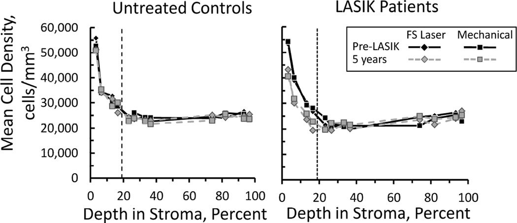
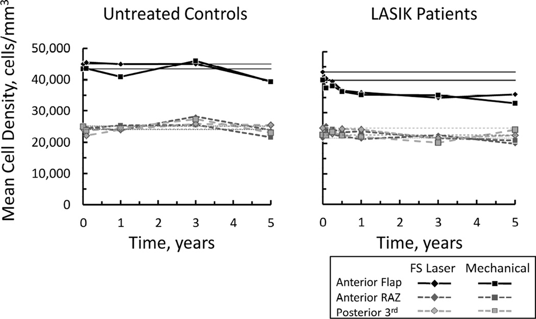
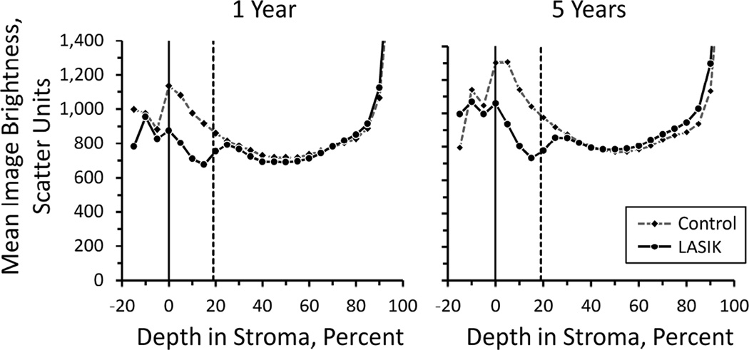
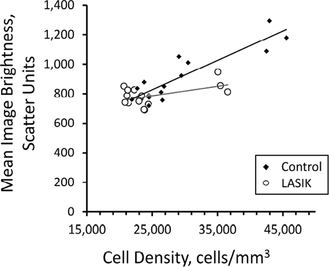
Results: Keratocyte density in the flap decreased by 20% during the first year after LASIK and remained low through 5 years (P < .001). High-order wavefront aberrations increased and uncorrected visual acuity improved immediately after surgery, but these variables did not change further to 5 years. There were no differences in any variables between treatments.
Conclusions: A sustained reduction in keratocyte density does not affect vision or optical properties of the cornea through 5 years after LASIK. The method of creating a LASIK flap does not influence the changes in keratocyte density in the flap.
Copyright © 2015 Elsevier Inc. All rights reserved.
Figures
References
-
- Erie JC, Nau CB, McLaren JW, Hodge DO, Bourne WM. Long-term keratocyte deficits in the corneal stroma after LASIK. Ophthalmology. 2004;111(7):1356–1361. - PubMed
-
- Pisella PJ, Auzerie O, Bokobza Y, Debbasch C, Baudouin C. Evaluation of corneal stromal changes in vivo after laser in situ keratomileusis with confocal microscopy. Ophthalmology. 2001;108(10):1744–1750. - PubMed
-
- Mitooka K, Ramirez M, Maguire LJ, et al. Keratocyte density of central human cornea after laser in situ keratomileusis. Am J Ophthalmol. 2002;133(3):307–314. - PubMed
-
- Canadas P, de Benito-Llopis L, Hernandez-Verdejo JL, Teus MA. Comparison of keratocyte density after femtosecond laser vs mechanical microkeratome from 3 months up to 5 years after LASIK. Graefes Arch Clin Exp Ophthalmol. 2013;251(9):2171–2179. - PubMed
Publication types
MeSH terms
Grants and funding
LinkOut - more resources
Full Text Sources
Other Literature Sources
