. 2015 Apr;14(4):357-68.
doi: 10.1111/gbb.12217.

The burrowing behavior of the nematode Caenorhabditis elegans: a new assay for the study of neuromuscular disorders

Affiliations

The burrowing behavior of the nematode Caenorhabditis elegans: a new assay for the study of neuromuscular disorders

C Beron et al. Genes Brain Behav. 2015 Apr.

Abstract

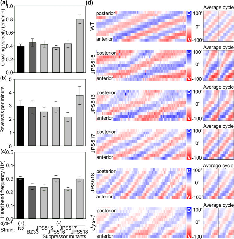
The nematode Caenorhabditis elegans has been a powerful model system for the study of key muscle genes relevant to human neuromuscular function and disorders. The behavioral robustness of C. elegans, however, has hindered its use in the study of certain neuromuscular disorders because many worm models of human disease show only subtle phenotypes while crawling. By contrast, in their natural habitat, C. elegans likely spends much of the time burrowing through the soil matrix. We developed a burrowing assay to challenge motor output by placing worms in agar-filled pipettes of increasing densities. We find that burrowing involves distinct kinematics and turning strategies from crawling that vary with the properties of the substrate. We show that mutants mimicking Duchenne muscular dystrophy by lacking a functional ortholog of the dystrophin protein, DYS-1, crawl normally but are severely impaired in burrowing. Muscular degeneration in the dys-1 mutant is hastened and exacerbated by burrowing, while wild type shows no such damage. To test whether neuromuscular integrity might be compensated genetically in the dys-1 mutant, we performed a genetic screen and isolated several suppressor mutants with proficient burrowing in a dys-1 mutant background. Further study of burrowing in C. elegans will enhance the study of diseases affecting neuromuscular integrity, and will provide insights into the natural behavior of this and other nematodes.

Keywords: Behavior; Caenorhabditis elegans; burrowing; dystrophin; nematode.


Conflict of interest statement

Conflict of interest: The authors declare no competing financial interests.

Figures

Figure 1
Figure 1
Kinematic analysis of the burrowing behavior of C. elegans. (a) We used 1.5-ml glass pipettes filled with agar of varying densities to film the burrowing behavior of individual worms. Adults were injected into one end of the pipette and filmed as they burrowed to an attractant (diacetyl) placed at the opposite end (inset). (b) Our custom algorithm implemented in ImagePro detected and digitized worms while freely behaving (top). The midline of animals were then divided into eleven sections of equal length (middle) and the angle between these was measured and assigned a color ranging from red for 70° ventral, to white for 0°, to blue for 70° dorsal. Here a burrowing worm is shown. Three color-coded frames are followed through the spinning process, which results in the creation of a behavioral matrix that describes the entire behavior of the animal over time (bottom). (c) A spined worm is shown (i) to illustrate the angles represented in the curvature matrix. A dorsal angle is presented as a shade of blue (ii), straight angles appear as white (iii), and ventral angles as shades of red (iv). In this and all curvature matrices, anterior is down and posterior is up.
Figure 2
Figure 2
Burrowing is distinct from swimming and crawling behaviors. (a) Burrowing head-bend frequency in a range of agar densities was lower than crawling frequency (mean and s.e.m. shown in green), and swimming (mean and s.e.m. shown in blue). N = 15 worms, 10 head-bends each, for swimming and crawling, and N = 10 worms, 10 head-bends each, for each agar density during burrowing. (b) Curvature matrix plot of a representative swimming bout 10 seconds long (i). The phase plot of matrix data averaged across individual head-bend cycles (ii) shows how the worm switches from a ventrally bent “C” shaped posture, pictured on the right (iii), to a dorsally bent C shaped posture and back. (c) Crawling worms moved by means of a persistent but propagating “S” shape (i), as depicted by the average cycle plot (ii) and picture (iii). (d) Across densities, worms burrowed with a continuous “M” (or “W”) shape. Burrowing is also distinct from swimming and crawling in lacking the dampening of posteriorly directed bends. All curvature matrices are ten seconds long. N = 10 worms with minimum of 10 cycles each for every condition, representative examples shown.
Figure 3
Figure 3
Worms alter their trajectory by different means depending on substrate properties. (a) Wild-type worms showed distinct strategies to change their direction depending on their environment. Swimming animals changed direction by producing deep bends and omega bends. Crawling worms favored reversals and burrowing worms rely on deep and lateral bends. (b) As substrate density increases, worms decreased their turning rate and relied increasingly on omega bends. At lower densities however worms used several turning strategies including lateral bends characterized by three-dimensional waves. Mean and s.e.m. reported for 3-min observation period after 30-min of acclimation for 30 worms in each condition.
Figure 4
Figure 4
Burrowing is an ideal behavior to assess neuromuscular integrity. We tested two dys-1 loss-of function alleles that model Duchenne muscular dystrophy (MD). Crawling behavior failed to produce striking phenotypes for these animals. For example, dys-1 worms display no defect in head-bend frequency (a), reversal rate (b), and velocity (c) when crawling. The same dys-1 mutant strains, however, were severely impaired in burrowing (d). Mean and s.e.m. reported for samples of 30 worms for the crawling, and 65 animals for the burrowing experiments. **P < 0.001 two-tailed t-test. (e) Unlike wild-type (top), burrowing dys-1 mutants had crawl-like kinematics (middle) interspersed with periods of immobility (bottom) as evident in the curvature matrices (10 sec each). Average curvature plots for corresponding strain on right.
Figure 5
Figure 5
Burrowing hastens muscular degeneration in worms modeling muscular dystrophy. Young (L4) wild-type and dys-1 mutant worms expressing GFP-tagged muscle nuclei and mitochondria (ccIs4251[Pmyo-3::GFP-NLS + Pmyo-3::GFP-mit]) were grown on either agar plates with bacteria, or in 6% agar pipettes with bacteria at opposite and half-way points to force animals to crawl or burrow respectively. After four days, crawling ability was tested and their musculature was subsequently imaged. Wild-type and dys-1 mutant worms showed only limited muscle degeneration when raised in the crawling condition ((a) top row); however, dys-1 mutants showed marked muscular degeneration when raised in the burrowing condition ((a) bottom row). Arrowheads point to areas of accumulation of GFP, indicative of muscle cell degeneration. Three representative worms are shown for each condition. The muscular degeneration observed for burrowing dys-1 mutants was reflected in their locomotor dysfunction. (b) dys-1(eg33) mutant worms raised in the burrowing condition showed a marked decrease in crawling velocity compared to their sisters raised in the crawling condition. (c) Although an increase in reversal frequency might be associated with the measured decrease in velocity for wild-type, this was not the case for dys-1 mutants. Instead, decreases in dys-1 crawling velocity seemed to be related to lower bending frequency (d). Here we report the mean and s.e.m. for N = 30 worms for each condition. ** P < 0.001 two-tailed t-test.
Figure 6
Figure 6
Suppression screen to identify mutations capable of rescuing the defective burrowing phenotype of worms modeling muscular dystrophy (dys-1(eg33) strain BZ33). (a) We devised a genetic screen to identify mutations capable of suppressing the poor burrowing ability of dys-1 mutant. A 2-μl drop of the attractant 1:100 diacetyl diluted in ethanol was placed on one side of a 3% agar-filled pipette. After 24 hrs worms were injected 20 cm away from the attractant and allowed to burrow within it. After 2 hours most wild-type worms will have migrated more than 4 cm toward the attractant (top). During the same time course, dys-1 mutants fail to make progress toward the attractant (middle). We mutagenized dys-1(eg33) worms and selected animals whose dys-1 phenotype had been suppressed by newly acquired mutations (bottom). (b) Four strains of suppressor mutants were isolated (JPS515, JPS516, JPS517 and JPS519) which displayed an improved ability to burrow over their dys-1 background.
Figure 7
Figure 7
Behavioral characterization of suppressor mutants. Like the dys-1(eg33) mutant strain BZ33, most suppressor mutants carrying the dys-1 mutation displayed wild-type crawling velocity (a), reversal rates (b), and head-bend frequencies (c). (d) We next compared their detailed burrowing kinematics to those of wild-type (top), and dys-1 mutant worms (bottom) with curvature matrices. Suppressor mutants had kinematics reminiscent of both wild-type and dys-1 worms. All bars show the mean and s.e.m. of 100 animals (in five trials) for burrowing, and 15 animals for crawling (30 for dys-1(eg33) vxJPS518). Burrowing bouts of 10 seconds in duration are shown, alongside average phase plots (right). Anterior is down, and posterior is up.
Figure 8
Figure 8
Suppressor mutation that improved burrowing ability also improved muscle integrity of the dys-1 mutant. Muscle degeneration that is normally exacerbated by burrowing conditions in the dys-1 mutant background was suppressed with the suppressor mutant strain JPS518. Note the wild-type-like pattern of GFP-labeled muscle mitochondria and nuclei from confocal stack images in both crawling and burrowing-raised conditions. Scale bar for all representative images on bottom.

References

    1. Bargmann CI, Horvitz HR. Chemosensory neurons with overlapping functions direct chemotaxis to multiple chemicals in C. elegans. Neuron. 1991;7:729–742. - PubMed
    1. Brenner S. The genetics of Caenorhabditis elegans. Genetics. 1974;77:71–94. - PMC - PubMed
    1. Croll NA. Components and patterns of in the behavior of the nematode Caenorhabditis elegans. J Zool. 1975;176:159–176.
    1. Culetto E, Sattelle DB. A role for Caenorhabditis elegans in understanding the function and interaction of human disease genes. Hum Mol Gen. 2000;9:869–877. - PubMed
    1. Davies KE, Nowak KJ. Molecular mechanisms of muscular dystrophies: Old and new players. Nat Rev Mol Cell Biol. 2006;7:762–773. - PubMed

Publication types

MeSH terms