. 2015 May 10;6(13):11561-74.
doi: 10.18632/oncotarget.3427.

Inhibition of β-catenin signaling suppresses pancreatic tumor growth by disrupting nuclear β-catenin/TCF-1 complex: critical role of STAT-3

Affiliations

Inhibition of β-catenin signaling suppresses pancreatic tumor growth by disrupting nuclear β-catenin/TCF-1 complex: critical role of STAT-3

Kartick C Pramanik et al. Oncotarget. .

Abstract

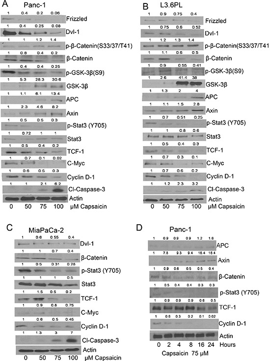
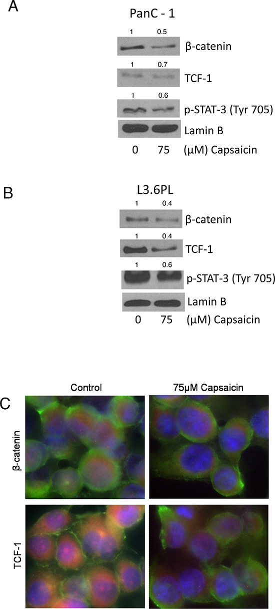
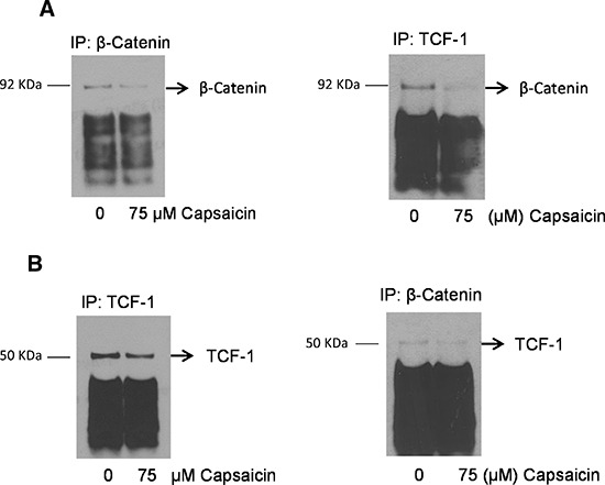
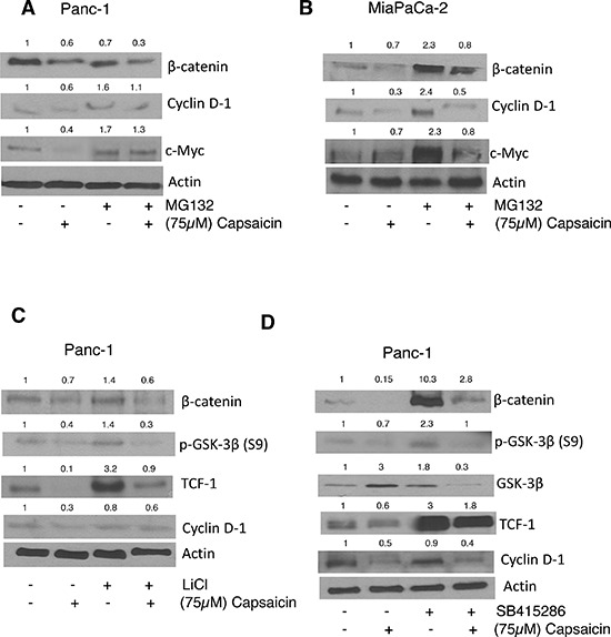
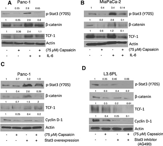
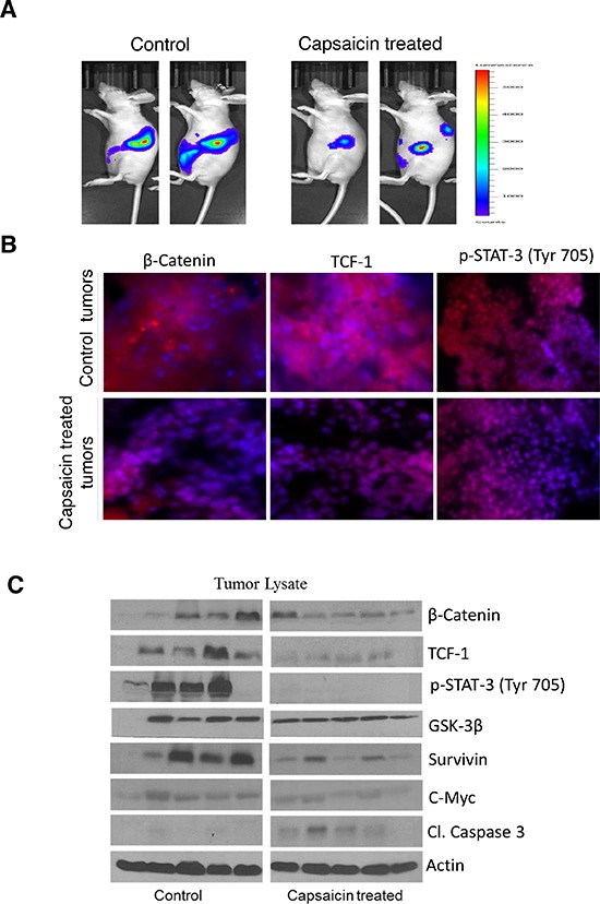
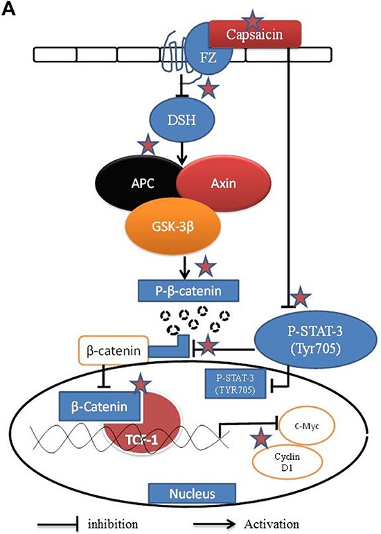
Aberrant activation of β-catenin/TCF signaling is related to the invasiveness of pancreatic cancer. In the present study, we evaluated the effect of capsaicin on β-catenin/TCF signaling. In a concentration and time-dependent study, we observed that capsaicin treatment inhibits the activation of dishevelled (Dsh) protein DvI-1 in L3.6PL, PanC-1 and MiaPaCa-2 pancreatic cancer cells. Capsaicin treatment induced GSK-3β by inhibiting its phosphorylation and further activated APC and Axin multicomplex, leading to the proteasomal degradation of β-catenin. Expression of TCF-1 and β-catenin-responsive proteins, c-Myc and cyclin D1 also decreased in response to capsaicin treatment. Pre-treatment of cells with MG-132 blocked capsaicin-mediated proteasomal degradation of β-catenin. To establish the involvement of β-catenin in capsaicin-induced apoptosis, cells were treated with LiCl or SB415286, inhibitors of GSK-3β. Our results reveal that capsaicin treatment suppressed LiCl or SB415286-mediated activation of β-catenin signaling. Our results further showed that capsaicin blocked nuclear translocation of β-catenin, TCF-1 and p-STAT-3 (Tyr705). The immunoprecipitation results indicated that capsaicin treatment reduced the interaction of β-catenin and TCF-1 in the nucleus. Moreover, capsaicin treatment significantly decreased the phosphorylation of STAT-3 at Tyr705. Interestingly, STAT-3 over expression or STAT-3 activation by IL-6, significantly increased the levels of β-catenin and attenuated the effects of capsaicin in inhibiting β-catenin signaling. Finally, capsaicin mediated inhibition of orthotopic tumor growth was associated with inhibition of β-catenin/TCF-1 signaling. Taken together, our results suggest that capsaicin-induced apoptosis in pancreatic cancer cells was associated with inhibition of β-catenin signaling due to the dissociation of β-catenin/TCF-1 complex and the process was orchestrated by STAT-3.

Keywords: GSK-3β; STAT3; orthotopic tumor; pancreatic cancer; β-catenin.


Conflict of interest statement

CONFLICT OF INTEREST

Authors declare no conflict of interest.

Figures

Figure 1
Figure 1. Effect of capsaicin on β-catenin and TCF-1 signaling pathway in pancreatic cancer cells
(A & D) PanC-1 (B) L3.63PL (C) MiaPaCa-2 cells were treated with various concentrations of capsaicin or at various time points with 75 μM of capsaicin for 24 h. Cells were lysed and subjected to western blot. Immunoblots were probed for Frizzled, DVI-1, p-β-catenin (S33/37/T41), β-catenin, p-GSK-3β (S9), GSK-3β, APC, Axin, p-Stat-3 (Tyr705), Stat-3, TCF-1, c-Myc, cyclin D1and Cl-caspase-3. Same blots were stripped and reprobed for actin to ensure equal protein loading. The experiments were repeated three times with similar results obtained.
Figure 2
Figure 2. Effect of capsaicin in nuclear localization of β-catenin, TCF-1 and p-Stat-3(Tyr 705)
(A) PanC-1 and (B) L3.6PL cells were treated with 75 μM capsaicin for 24 h and nuclear fraction were isolated using nuclear fractionation kit. Represented western blots were immunoblotted with β-catenin, TCF-1 and p-Stat-3 (Tyr705) antibodies. The same blots were then stripped and reprobed with lamin B to ensure equal protein loading. (C) PanC-1 cells were treated with DMSO or 75 μM capsaicin, immunostained with β-catenin and TCF-1 (red), and actin (green) antibodies, then visualized under fluorescence microscope (Olympus Inc.). The experiments were repeated three times with similar results obtained.
Figure 3
Figure 3. Capsaicin treatment disrupts nuclear β-catenin and TCF-1 complex
PanC-1 cells were treated with DMSO or 75 μM capsaicin for 24 h and nuclear fraction was isolated by nuclear fractionation kit and immunoprecipitated with (A & B) β-catenin and TCF-1 and immunobloted with β-catenin and TCF-1 antibodies respectively. The experiments were repeated three times with similar results obtained.
Figure 4
Figure 4. Effect of capsaicin on MG132, Licl and SB415286 treatment
(A) PanC-1 and (B) MiaPaCa-2 cells were pre-treated with 10 μM MG-132 followed by treatment with 75 μM capsaicin for 24 h. After which whole cell lysates were immunoblotted with β-catenin, cyclin D1, c-Myc (C&D) in a separate experiment, PanC-1 cells were pre-treated with 40 mM Licl or 50 μM SB415286, followed by 75 μM capsaicin treatment for 24 h and then whole cell lysates were immunoblotted with β-catenin, p-GSK-3β, GSK-3β, TCF-1, Cyclin D1. The same blot was stripped and reprobed for actin to ensure equal protein loading.
Figure 5
Figure 5. Effect of capsaicin on IL-6, Stat-3 inhibitor and Stat-3 over-expression plasmid
(A) PanC-1 (B) MiaPaCa-2 cells were Pre-treated with 20 ng/ml IL-6 followed by 75 μM capsaicin treatment for 24 h. (C) PanC-1 cells were transiently transfected with 2 μg of Stat-3 plasmid using FuGENE transfection reagent for 24 h and then transfected cells were treated with 75 μM capsaicin for 24 h. (D) L3.6PL cell were pre-treated with 10 μM Stat-3 inhibitor, followed by 75 μM capsaicin treatment for 24 h. Whole cell lysates were immunobloted with p-Stat-3 (Tyr 705), β-catenin, TCF-1, Cyclin D1. The same blots were stripped and reprobed for actin to ensure equal protein loading.
Figure 6
Figure 6. Capsaicin inhibits the growth of orthotropic pancreatic tumor by inhibiting β-catenin/TCF-1 signaling
In the present study we used our previous orthotopic experiment tumors to represent the following parameters. (A) Around 1 × 10⁶ PanC-1-luc cells were injected orthotopically in the pancreas with minor surgery. Once mice had stable image, animals were randomly divided into two groups. The treated group received 5 mg/kg body weight capsaicin by oral gavage every day, whereas control group received vehicle only. Animals were imaged using IVIS Bio Luminescent System. Representative images of control and capsaicin treated mice are shown in Figure 6A. (B) Tumors from control and capsaicin treated mice were dissected out and kept in 4% formalin solution. Tumors were then sliced about 10 μm thick and placed on glass slides and kept in frozen for 24 h. Treated and untreated tumors were immunostained with β-catenin (red), TCF-1(red) and p-Stat-3 (Tyr 705) (red) antibodies and visualized under fluorescence microscope (Olympus Inc.). The experiments were repeated three times with similar results obtained. In order to determine the mechanism of tumor growth suppression, tumors were homogenized, lysed and subjected to western blot. (C) Representative immunnoblots showed the effect of capsaicin treatment on phosphorylation of p-Stat-3 (Tyr 705) and protein levels of β-catenin, TCF-1, GSK-3β, Survivin, c-Myc and Cl-caspase-3. Each band represents tumor from different mouse. The blots were stripped and reprobed for actin to ensure equal protein loading.
Figure 7
Figure 7. Schematic mechanism of action of capsaicin in pancreatic cancer cells targeting β-catenin pathway
☆Preferential sites of action by capsaicin.

References

    1. Li J, Wientjes MG, Au JL. Pancreatic cancer: pathobiology, treatment options, and drug delivery. The AAPS Journal. 2010;12:223–232. - PMC - PubMed
    1. Heiser PW, Cano DA, Landsman L, Kim GE, Kench JG, Klimstra DS, Taketo MM, Biankin AV, Hebrok M. Stabilization of beta-catenin induces pancreas tumor formation. Gastroenterology. 2008;135:1288–1300. - PMC - PubMed
    1. Zeng G, Germinaro M, Micsenyi A, Monga NK, Bell A, Sood A, Malhotra V, Sood N, Midda V, Monga DK, Kokkinakis DM, Monga SP. Aberrant Wnt/beta-catenin signaling in pancreatic adenocarcinoma. Neoplasia. 2006;8:279–289. - PMC - PubMed
    1. Wang L, Heidt DG, Lee CJ, Yang H, Logsdon CD, Zhang L, Fearon ER, Ljungman M, Simeone DM. Oncogenic function of ATDC in pancreatic cancer through Wnt pathway activation and beta-catenin stabilization. Cancer Cell. 2009;15:207–219. - PMC - PubMed
    1. Polakis P. Wnt signaling and cancer. Genes & development. 2000;14:1837–1851. - PubMed

Publication types

MeSH terms