. 2015 May 20;6(14):12587-602.
doi: 10.18632/oncotarget.3494.

Myc-dependent purine biosynthesis affects nucleolar stress and therapy response in prostate cancer

Affiliations

Myc-dependent purine biosynthesis affects nucleolar stress and therapy response in prostate cancer

Stefan J Barfeld et al. Oncotarget. .

Abstract

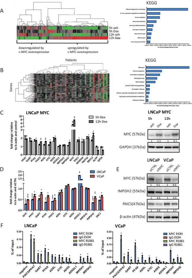
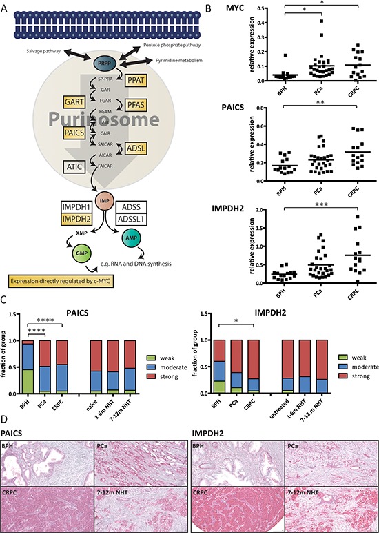
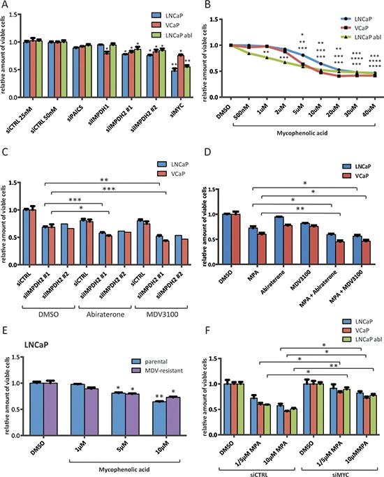
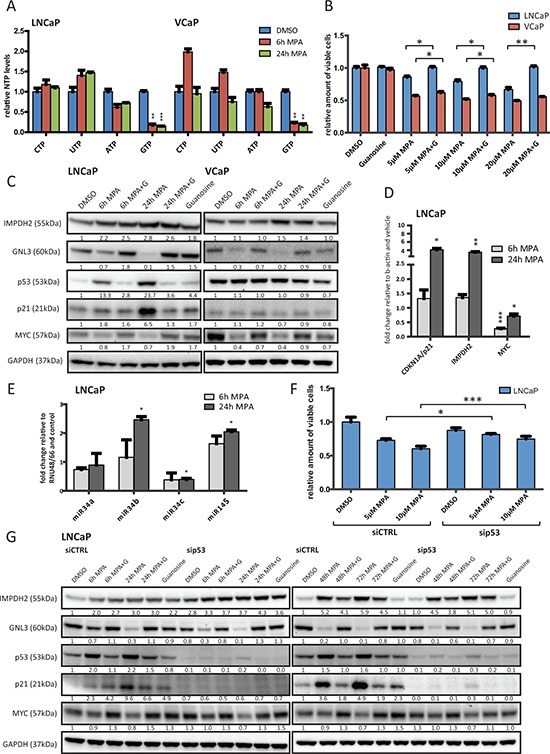
The androgen receptor is a key transcription factor contributing to the development of all stages of prostate cancer (PCa). In addition, other transcription factors have been associated with poor prognosis in PCa, amongst which c-Myc (MYC) is a well-established oncogene in many other cancers. We have previously reported that the AR promotes glycolysis and anabolic metabolism; many of these metabolic pathways are also MYC-regulated in other cancers. In this study, we report that in PCa cells de novo purine biosynthesis and the subsequent conversion to XMP is tightly regulated by MYC and independent of AR activity. We characterized two enzymes, PAICS and IMPDH2, within the pathway as PCa biomarkers in tissue samples and report increased efficacy of established anti-androgens in combination with a clinically approved IMPDH inhibitor, mycophenolic acid (MPA). Treatment with MPA led to a significant reduction in cellular guanosine triphosphate (GTP) levels accompanied by nucleolar stress and p53 stabilization. In conclusion, targeting purine biosynthesis provides an opportunity to perturb PCa metabolism and enhance tumour suppressive stress responses.

Keywords: cancer; metabolism; nucleotide; prostate; transcription.


Figures

Figure 1
Figure 1. The expression of purine biosynthesis enzymes in prostate cancer cells is directly regulated by MYC
A. Illumina beadarray results. LNCaP MYC cells were hormone-deprived for 72 h prior to stimulation with 2 μg/ml Doxycycline or vehicle for the indicated time points to induce MYC-overexpression. Total RNA of three independent experiments was isolated and subjected to expression array analysis. Genes with significantly altered expression (q < 0.05) compared to vehicle treated cells are displayed using unsupervised hierarchical clustering (left). Functional annotation using KEGG pathway analysis of 266 genes defined as ‘upregulated by MYC-overexpression at both time points’ is shown (right). B. Expression data from a previously published study (Taylor et al.) was used to define candidate MYC-regulated genes. Only patients marked as ‘biochemical recurrence (BCR)’ or ‘metastases (METS)’ were included in the analysis. Similarity search using Pearson correlation (1% tolerance) was performed on MYC expression and displayed using unsupervised hierarchical clustering with genes on the Y-axis and patients on the X-axis (left). Functional annotation of the 264 MYC-correlated genes using KEGG pathway analysis at 1% tolerance is shown (right) C. Real-time PCR results of MYC-overexpressing cells. LNCaP MYC cells were hormone-deprived for 72 h prior to stimulation with 2 μg/ml Doxycycline to induce MYC-overexpression. Total RNA was isolated, reverse transcribed and used for qRT-PCR. n = 3 (D-E) Cells were transfected with 50 nM of control or MYC siRNA for 72 h. D. Real-time PCR results of MYC-depleted cells. Total RNA was isolated, reverse transcribed and used for qRT-PCR. n = 3–4 E. Western Blot results of MYC-depleted cells. Protein lysates were harvested, separated by SDS-PAGE and blotted for the indicated proteins. Protein levels were normalized to siCTRL and β-actin levels. Densitometry analysis of three biological replicates with SEM can be found in Figure S1H. F. ChIP-qPCR detection of promoter regions of purine biosynthesis genes in MYC ChIP samples. Cells were hormone-deprived for 72 h prior to stimulation with 1 nM R1881 or vehicle for 4 h. Subsequently, chromatin was crosslinked, sonicated and subjected to ChIP using an antibody against MYC or an unspecific IgG control. n = 3.
Figure 2
Figure 2. PAICS and IMPDH2 are overexpressed in prostate cancer patients
A. Schematic overview of the de novo purine biosynthesis pathway with direct MYC targets highlighted in yellow. B. Real-time PCR results on clinical samples. Total RNA was collected from prostate biopsies of patients suffering from benign hyperplasia (BPH), prostate cancer (PCa) or castrate-resistant prostate cancer (CRPC). The expression levels of MYC, PAICS and IMPDH2 were measured using real-time PCR and normalized to the expression of TATA-box binding protein (TBP). The line displays the mean value and significance was determined using one-way analysis of variation (ANOVA) with Bonferroni's multiple comparison test. n = 15–27 C. Immunohistochemistry data. Staining intensities of PAICS and IMPDH2 in patient biopsies with BPH, PCa CRPC, hormone-naïve, short and long NHT were assessed. Intensities divided into four groups (negative = 0, weak = 1, moderate = 2 and strong = 3) and used for subsequent analysis. Statistical analysis was performed using one-way analysis of variation (ANOVA) with Bonferroni's multiple comparison test. n = 20–113 cores D. Representative images of the TMAs for PAICS (left) and IMPDH2 (right).
Figure 3
Figure 3. Inhibition of IMPDH2 impairs the proliferation of prostate cancer cells and shows additive effects with established anti-androgens
A. Cell viability results of siRNA treated cells. Cells were transfected with 25 nM (control 25 nM, IMPDH1 and IMPDH2 #1) or 50 nM (control 50 nM, PAICS, IMPDH2 #2 and MYC) for 72 h and cell viability relative to siCTRL 50 nM was determined using a MTS-based assay. n = 2–4 B. Cell viability results of MPA treated cells. Cells were allowed to attach for 48 h prior to treatment with indicated doses of MPA for 72 h. Cell viability relative to vehicle control was determined using a MTS-based assay. n = 2–4 C. Cell viability results of siRNA and Abiraterone/MDV3100 treated cells. Cells were transfected with 25 nM IMPDH2 #1, 50 nM IMPDH2 #2 siRNA or equal amounts of siCTRL for 48 h. Following treatment with the indicated drugs for another 72 h, viability relative to DMSO and siCTRL was assessed using a MTS-based assay. Doses for LNCaP were 1 μM Abiraterone and 1 μM MDV3100, and for VCaP 1 μM Abiraterone and 100 nM MDV3100 n = 1–4 D. Cell viability results of MPA and Abiraterone/MDV3100 treated cells. Cells were allowed to attach for 48 h prior to treatment with indicated drug combinations for 72 h. Cell viability relative to vehicle control was determined using a MTS-based assay. Doses for LNCaP were 10 μM MPA, 1 μM Abiraterone and 1 μM MDV3100, and for VCaP 5 μM MPA, 1 μM Abiraterone and 100 nM MDV3100 n = 3–4 E. Cell viability results of MPA treated parental and MDV3100-resistant LNCaP. Cells were allowed to attach for 48 h prior to treatment with the indicated doses of MPA for 72 h. Cell viability relative to vehicle control was determined using a MTS-based assay. n = 4 F. Cell viability results of siRNA and MPA treated cells. Cells were transfected with 50 nM MYC or control siRNA for 48 h. Following treatment with the indicated doses of MPA (LNCaP 5 and 10 μM, VCaP 5 and 10 μM and LNCaP abl 1 and 10 μM) for another 72 h, viability was assessed using a MTS-based assay and normalized to the respective DMSO control. n = 3.
Figure 4
Figure 4. Inhibition of IMPDH2 leads to nucleolar instability, p53 activation and upregulation of MYC-targeting miRs
A. HPLC results of cellular nucleoside triphosphate levels. LNCaP and VCaP cells were treated with 10 μM MPA for the indicated time points, lysed and subjected to HPLC to detect cellular levels of presented nucleoside triphosphates (NTP). Values were normalized to DMSO treated cells and protein content. n = 3 B. Cell viability results of MPA treated cells. Cells were allowed to attach for 48 h prior to treatment with indicated doses of MPA and guanosine (100 μM) for 72 h. Cell viability relative to vehicle control was determined using a MTS-based assay. n = 3 C. Western blot results of MPA treated PCa cells. LNCaP and VCaP cells were treated with 10 μM MPA and 100 μM guanosine (G) for the indicated time points. Protein extracts were harvested and subjected to Western blot analysis using the indicated primary antibodies. Protein levels were normalized to DMSO controls and GAPDH levels. D. Real-time PCR results. LNCaP cells were treated with 10 μM MPA for the indicated time points. Total RNA was isolated, reverse transcribed and used for qRT-PCR. n = 3 E. miRNA detection using real-time PCR of MPA treated LNCaP. Cells were treated with 10 μM MPA for the indicated time points prior to total RNA isolation using Trizol reagent and miR-detection using TaqMan assays. n = 2 F. Cell viability results of siRNA and MPA treated cells. Cells were transfected with 50 nM control or p53 siRNA for 48 h. Following treatment with the indicated drugs for another 72 h, viability was assessed using a MTS-based assay. n = 3 G. Western Blot results of siRNA and MPA treated LNCaP. Cells were transfected with 50 nM control or p53 siRNA for 48 h and then treated with 10 μM MPA and 100 μM guanosine (G) for the indicated time points. Protein lysates were harvested, separated by SDS-PAGE and blotted for the indicated proteins.

References

    1. Denmeade SR, Isaacs JT. A history of prostate cancer treatment. Nat Rev Cancer. 2002;2:389–396. - PMC - PubMed
    1. Feldman BJ, Feldman D. The development of androgen-independent prostate cancer. Nat Rev Cancer. 2001;1:34–45. - PubMed
    1. van der Kwast TH, Schalken J, Ruizeveld de Winter JA, van Vroonhoven CC, Mulder E, Boersma W, Trapman J. Androgen receptors in endocrine-therapy-resistant human prostate cancer. Int J Cancer. 1991;48:189–193. - PubMed
    1. Sadi MV, Walsh PC, Barrack ER. Immunohistochemical study of androgen receptors in metastatic prostate cancer. Comparison of receptor content and response to hormonal therapy. Cancer. 1991;67:3057–3064. - PubMed
    1. Koivisto P, Kononen J, Palmberg C, Tammela T, Hyytinen E, Isola J, Trapman J, Cleutjens K, Noordzij A, Visakorpi T, Kallioniemi OP. Androgen receptor gene amplification: a possible molecular mechanism for androgen deprivation therapy failure in prostate cancer. Cancer Res. 1997;57:314–319. - PubMed

Publication types

MeSH terms