. 2015 Oct;35(7):943-52.
doi: 10.1007/s10571-015-0189-3. Epub 2015 Apr 14.

Ginkgolide B Protects Neurons from Ischemic Injury by Inhibiting the Expression of RTP801

Affiliations

Ginkgolide B Protects Neurons from Ischemic Injury by Inhibiting the Expression of RTP801

Xiaomei Wu et al. Cell Mol Neurobiol. 2015 Oct.

Abstract

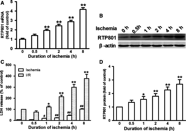
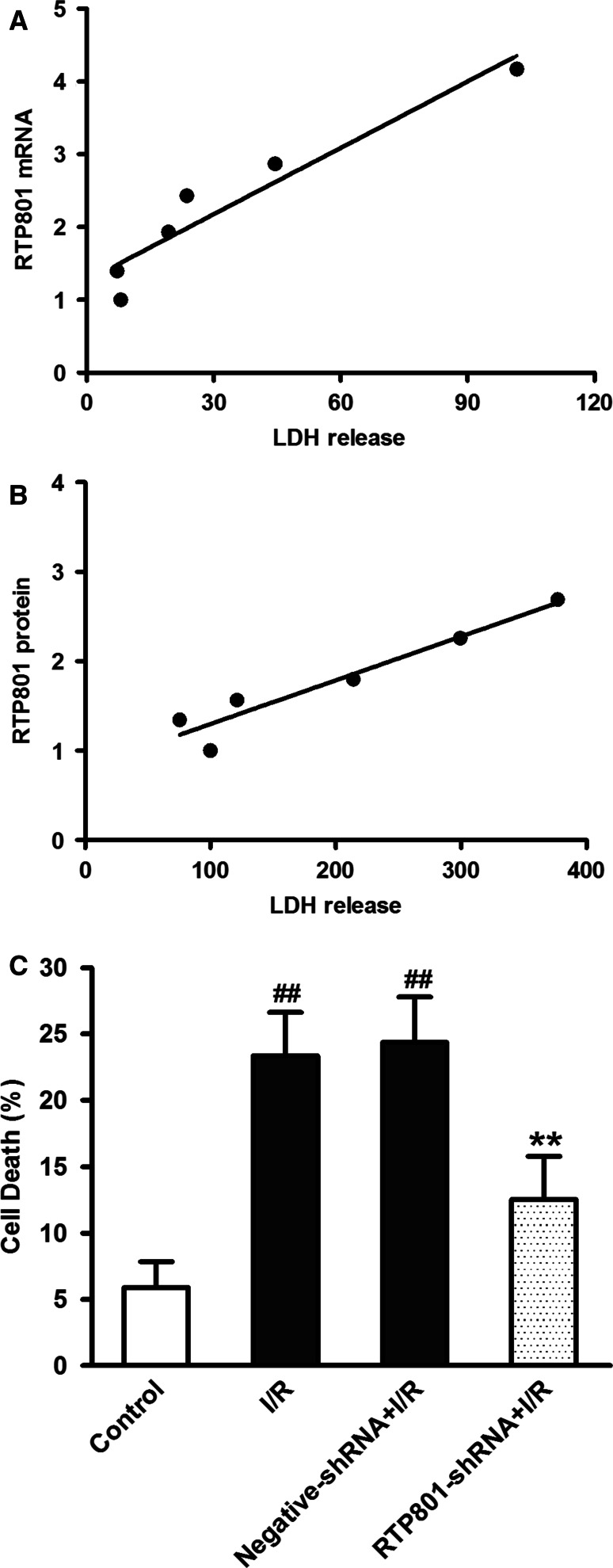
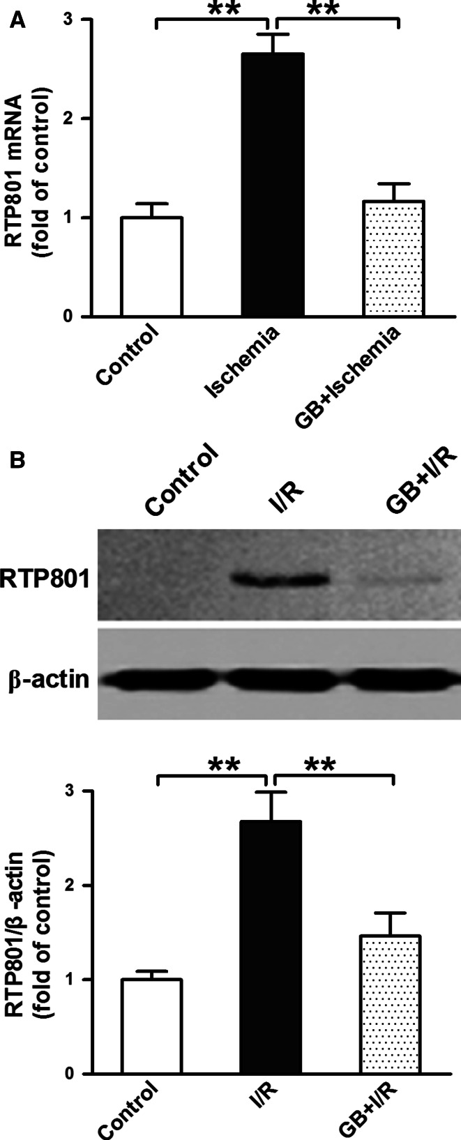
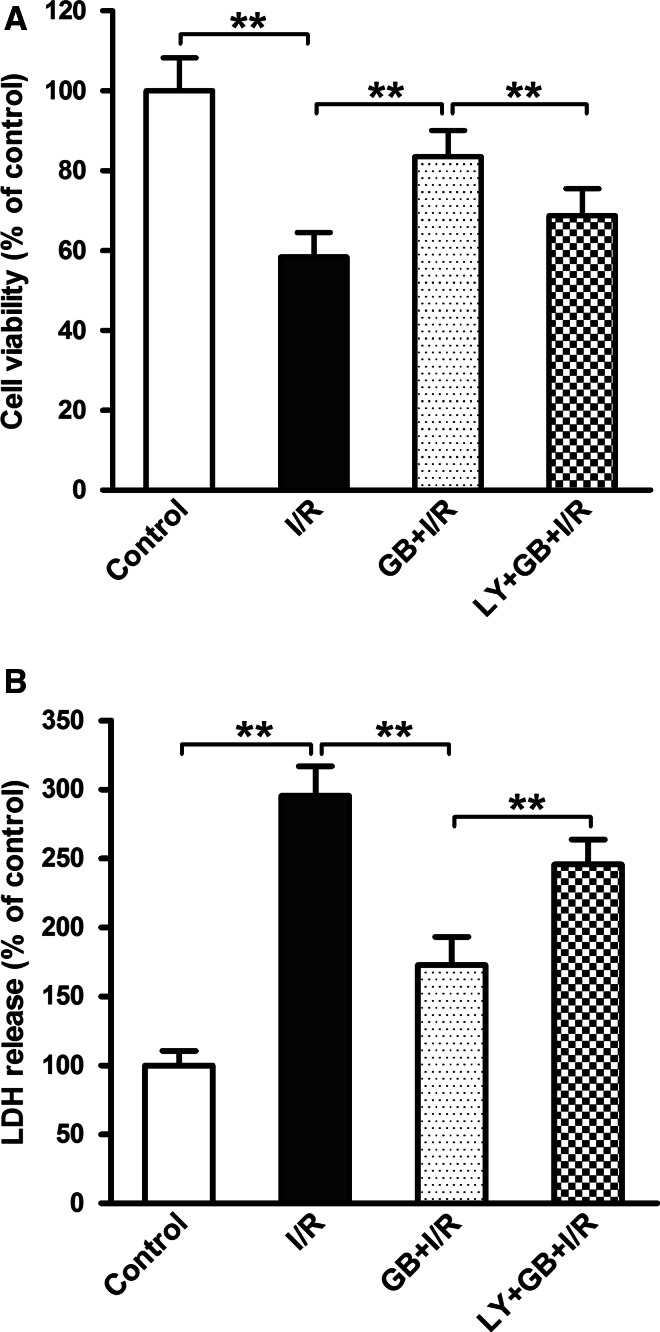
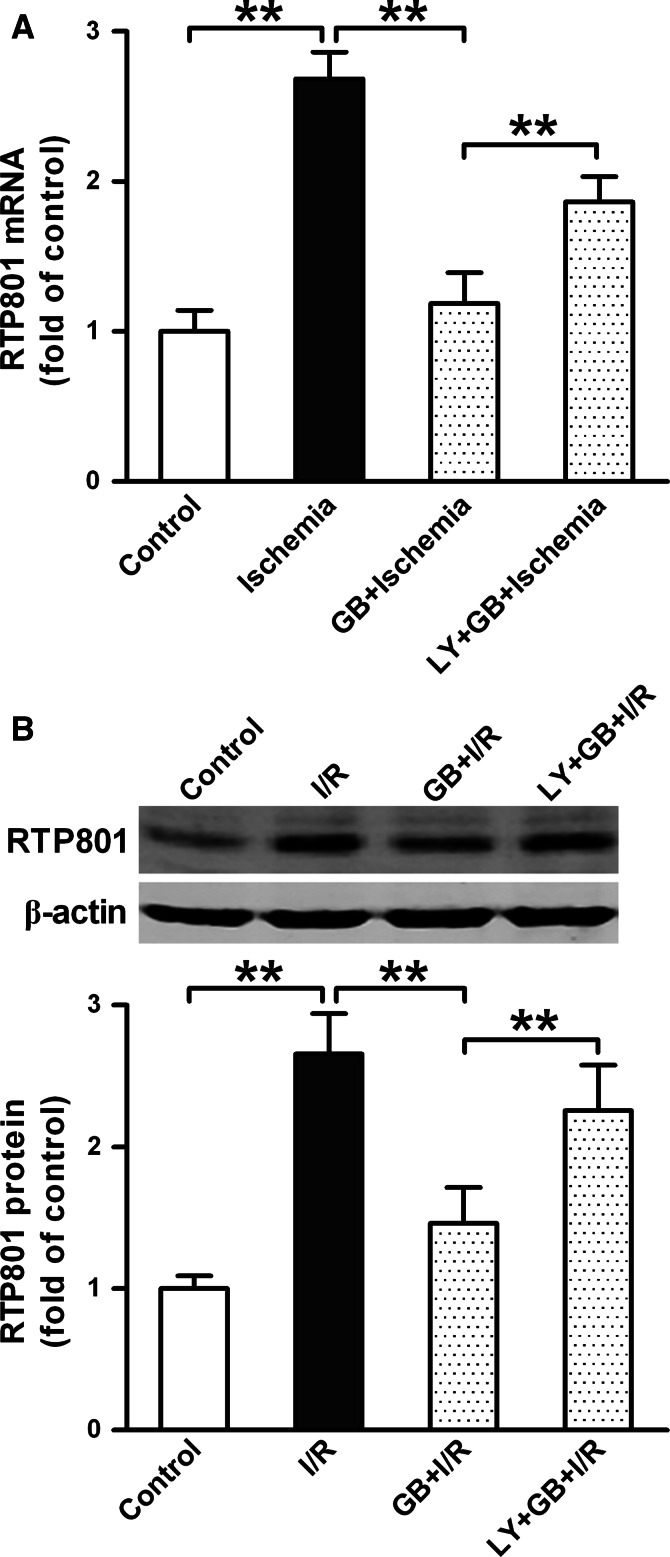
RTP801 (also known as REDD1), a stress-related protein, is induced by several environmental stresses such as ischemia and cigarette smoke. Although ischemia can dramatically up-regulate RTP801 expression in brain ischemia, up to now, the exact relation between RTP801 and neuronal death in ischemia is poorly understood. In the current study, using oxygen and glucose deprivation as an in vitro ischemic model in primary cultured cortical neurons, we found that the expression of RTP801 increased progressively with prolongation of ischemic duration, in which the expression of RTP801 is positively correlated with the release of lactate dehydrogenase (LDH) in neurons, and knockdown of RTP801 promoted neuronal survival in ischemia-reperfusion. It was further found that ginkgolide B (GB) could significantly increase cell viability and decrease LDH release, and at the same time reduce the levels of RTP801 mRNA and protein in neurons after ischemia and reperfusion. Moreover, GB-induced reduction in expression of RTP801 was blocked by application of LY294002, a specific inhibitor of phosphatidylinositol 3-kinase (PI3K). These results demonstrate that RTP801 could play a detrimental role on neurons in ischemia, and GB might protect neurons against ischemic injury by inhibiting RTP801 expression via PI3K pathway.

Keywords: Ginkgolide B; Ischemia; Ischemia–Reperfusion; Neuron; Phosphatidylinositol 3-kinase; RTP801.


Conflict of interest statement

The authors state no conflict of interest.

Figures

Fig. 1
Fig. 1
Effects of ischemia for different periods on cell viability and RTP801 expression in neurons. Neurons were subjected to oxygen and glucose deprivation (OGD) for 0, 0.5, 1, 2, 4, or 8 h followed or not by returning the cells to normal medium and reoxygenation for 24 h. RTP801 expression in neurons was detected by real-time RCR and Western blot methods, and the cell viability of neurons was measured after ischemia or ischemia–reperfusion (I/R) by LDH assay. a The expression of RTP801 mRNA in neurons after ischemia. b A representative Western blot of RTP801 in neurons after ischemia–reperfusion (I/R). c Quantification of expression of RTP801 (n = 3). d Cell viabilities of neurons after ischemia and ischemia–reperfusion (the percentage of control in I/R, n = 6). The data were analyzed by one-way ANOVA followed by Newman–Keuls post hoc test and presented as Mean ± SD. # p < 0.05, ## p < 0.01 versus 0 h in ischemic condition; *p < 0.05, **p < 0.01 versus 0 h in I/R condition
Fig. 2
Fig. 2
Relationship between RTP801 and neuronal damage under ischemic condition. The Pearson correlation between the expression of RTP801 and the release of LDH in the neurons treated with ischemia for different durations was observed by plotting the values for these two indicators against one another. a Relationship between the levels of RTP801 mRNA and the release of LDH after treatment with ischemia. b Relationship between the levels of RTP801 protein and the release of LDH after treatment with ischemia–reperfusion. c Effect of RTP801 knockdown on neuronal death in ischemia–reperfusion. Neurons were infected with lentivirus expressing GFP and shRNA for RTP801 for 72 h before being subjected to ischemia–reperfusion. Neuronal death was detected by trypan blue exclusion test. The percentage of neuronal death was determined by counting the number of double-positive (GFP+/trypan blue+) cells in total GFP-positive cells per field (40× objective) in 6 different fields per well. The data were analyzed by two-way ANOVA followed by Newman–Keuls post hoc test and presented as Mean ± SD. ## p < 0.01 versus control; **p < 0.01 versus negative-shRNA in I/R
Fig. 3
Fig. 3
Effects of ginkgolide B on the expression of RTP801 in neurons during ischemia and reperfusion. Neurons were pre-treated with GB at a concentration of 120 μmol/L for 24 h before being exposed to ischemia and I/R. The expression of RTP801 mRNA was measured by real-time RT-PCR after ischemia, and the level of RTP801 protein was detected by Western blot method after I/R. a The expression of RTP801 mRNA in neurons. b The expression of RTP801 protein in neurons. Statistical significance was assessed by two-way ANOVA followed by a Newman–Keuls post hoc test. Data were presented as Mean ± SD (n = 3). **p < 0.01
Fig. 4
Fig. 4
Effect of inhibitor of PI3K on the protection by ginkgolide B on neurons in ischemia–reperfusion. Neurons were treated with GB at 120 μmol/L for 24 h before being exposed to ischemia for 4 h followed by 24-h reperfusion. Sister neurons were pre-treated with LY294002 (LY, a specific inhibitor of PI3K, 50 μmol/L) for 1 h before being treated with GB and followed by ischemia–reperfusion. The cell viability of neurons was measured after I/R by MTT method (a) and LDH assay (b). Statistical significance was assessed by two-way ANOVA followed by a Newman–Keuls post hoc test. Data were expressed as a percentage of control (mean ± SD, n = 8). **p < 0.01
Fig. 5
Fig. 5
Effect of inhibitor of PI3K on the expression of RTP801 in neurons pre-treated with ginkgolide B in ischemia–reperfusion. Neurons were pre-treated with LY294002 (LY, a specific inhibitor of PI3K, 50 μmol/L) for 1 h before being treated with GB (120 μmol/L) for 24 h and then subjected to ischemia–reperfusion. RTP801 mRNA was measured after ischemia by real-time RT-PCR, and the level of RTP801 protein was detected after I/R by Western blot method. a The expression of RTP801 mRNA in neurons. b The expression of RTP801 protein in neurons. Statistical significance was assessed by two-way ANOVA followed by a Newman–Keuls post hoc test. Data were presented as Mean ± SD (n = 3). **p < 0.01

Similar articles

Cited by

References

    1. Bate C, Kempster S, Williams A (2006) Platelet-activating factor antagonists protect amyloid-beta damaged neurons from microglia-mediated death. Neuropharmacology 51(2):173–181 - PubMed
    1. Brugarolas J, Lei K, Hurley RL, Manning BD, Reiling JH, Hafen E, Witters LA, Ellisen LW, Kaelin WG (2004) Regulation of mTOR function in response to hypoxia by REDD1 and the TSC1/TSC2 tumor suppressor complex. Genes Dev 18(23):2893–2904 - PMC - PubMed
    1. Chung HS, Harris A, Kristinsson JK, Ciulla TA, Kagemann C, Ritch R (1999) Ginkgo biloba extract increases ocular blood flow velocity. J Ocul Pharmacol Ther 15(3):233–240 - PubMed
    1. Fang D, Li Z, Zhong-ming Q, Mei WX, Ho YW, Yuan XW, Ya K (2008) Expression of bystin in reactive astrocytes induced by ischemia/reperfusion and chemical hypoxia in vitro. Biochim Biophys Acta 1782(11):658–663 - PubMed
    1. Fang W, Deng Y, Li Y, Shang E, Fang F, Lv P, Bai L, Qi Y, Yan F, Mao L (2010) Blood brain barrier permeability and therapeutic time window of ginkgolide B in ischemia–reperfusion injury. Eur J Pharm Sci 39(1–3):8–14 - PubMed

Publication types

MeSH terms

LinkOut - more resources