Review
. 2015 Jun:83:1-13.
doi: 10.1016/j.yjmcc.2015.04.005. Epub 2015 Apr 11.

So! What's aging? Is cardiovascular aging a disease?

Affiliations
Review

So! What's aging? Is cardiovascular aging a disease?

Edward G Lakatta. J Mol Cell Cardiol. 2015 Jun.

Abstract

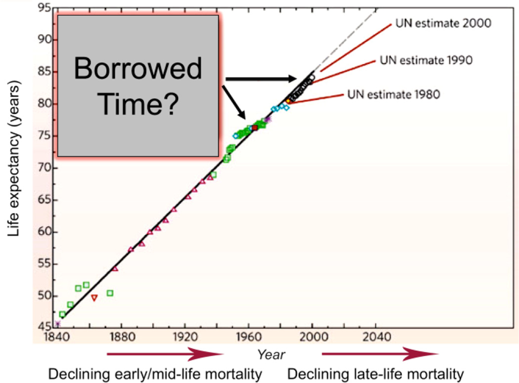
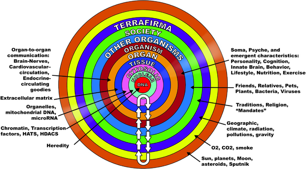
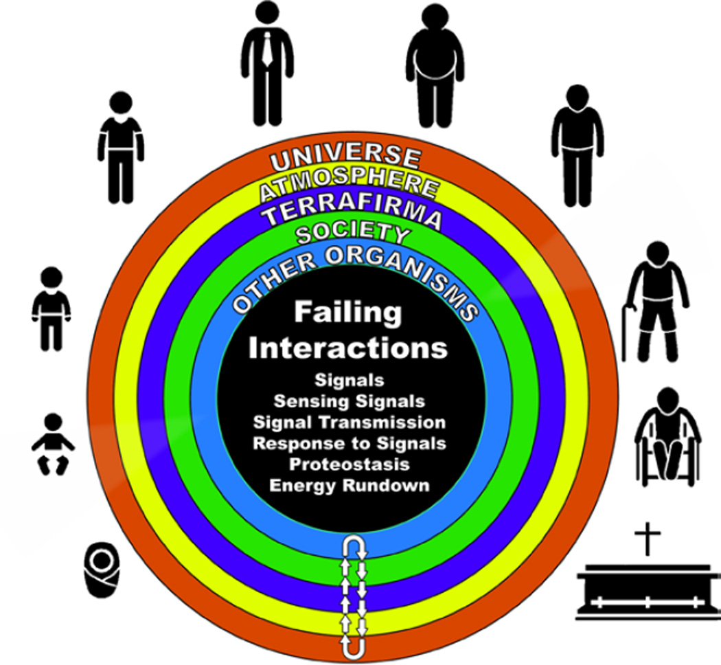
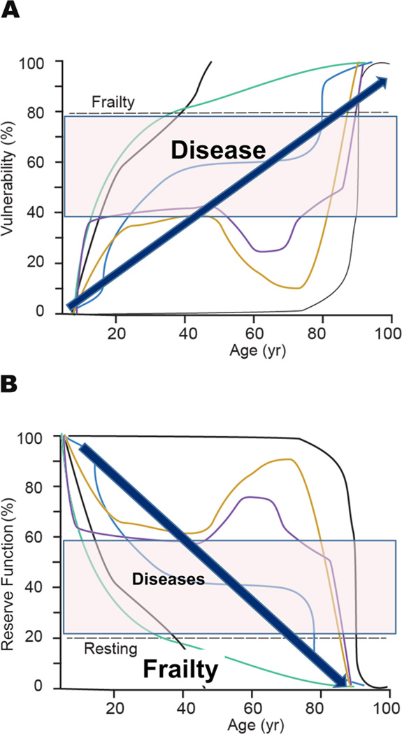
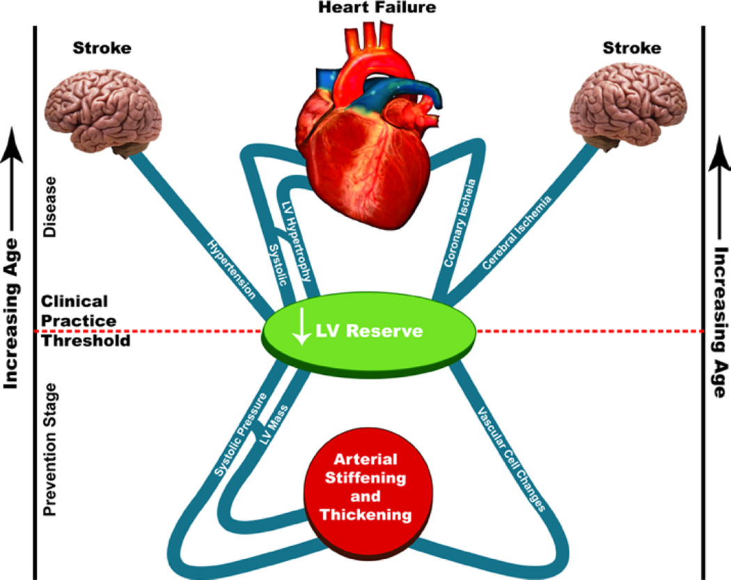
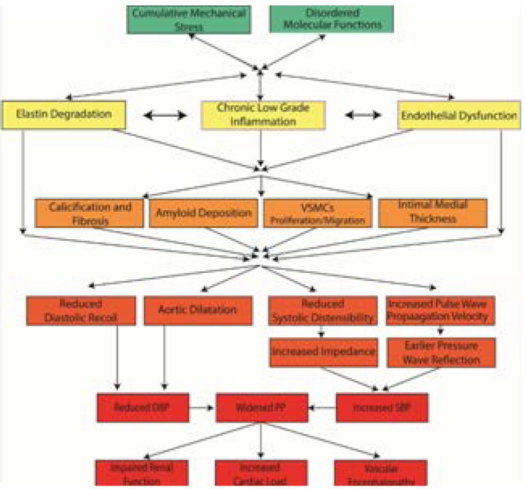
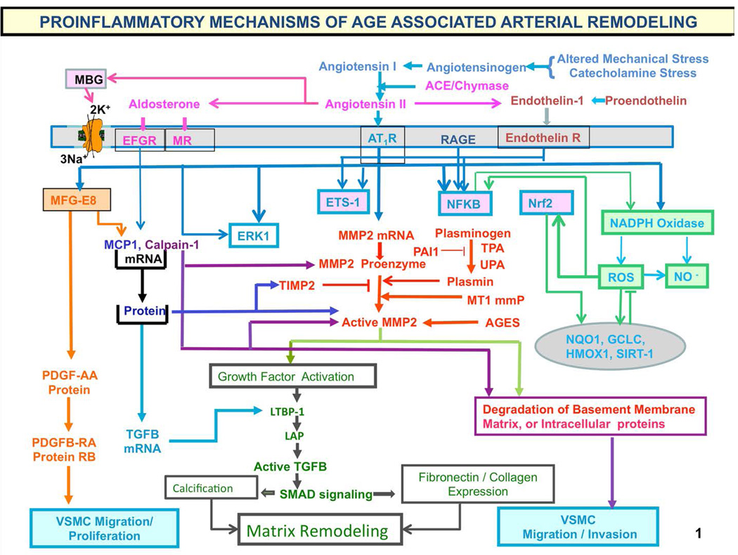
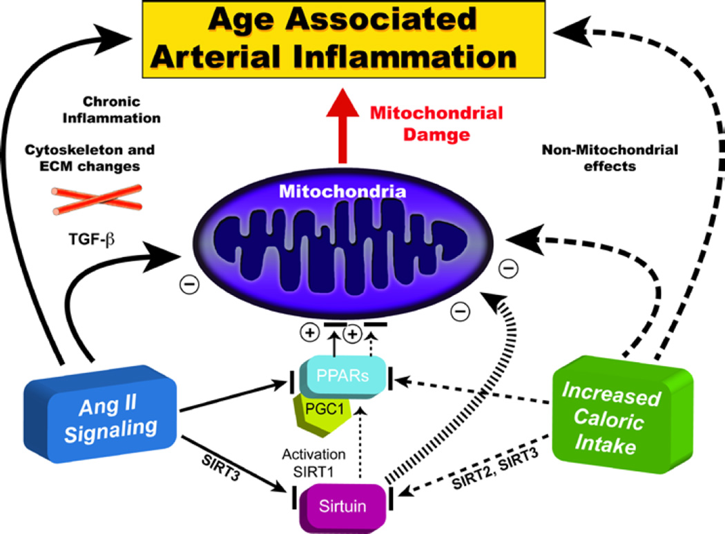
"Inside every old person is a young person wondering what happened." So, what is aging? Aging is a manifestation of progressive, time-dependent failure of molecular mechanisms that create disorder within a system of DNA and its environment (nuclear, cytosolic, tissue, organ, organism, other organisms, society, terra firma, atmosphere, universe). Continuous signaling, transmitted with different kinetics across each of these environments, confers a "mutual enslavement" that creates ordered functions among the components within the system. Accrual of this molecular disorder over time, i.e. during aging, causes progressive changes in the structure and function of the heart and arteries that are quite similar in humans, non-human primates, rabbits and rats that compromise cardiovascular reserve function, and confer a marked risk for incident cardiovascular disease. Nearly all aspects of signaling within the DNA environment system within the heart and arteries become disordered with advancing age: Signals change, as does sensing of the signals, transmission of signals and responses to signals, impaired cell renewal, changes in the proteome due to alterations in genomic transcription, mRNA translation, and proteostasis. The density of some molecules becomes reduced, and post-translational modifications, e.g. oxidation and nitration phosphorylation, lead to altered misfolding and disordered molecular interactions. The stoichiometry and kinetics of enzymatic and those reactions which underlie crucial cardiac and vascular cell functions and robust reserve mechanisms that remove damaged organelles and proteins deteriorate. The CV cells generate an inflammatory defense in an attempt to limit the molecular disorder. The resultant proinflammatory milieu is not executed by "professional" inflammatory cells (i.e. white blood cells), however, but by activation of renin-angiotensin-aldosterone endothelin signaling cascades that leads to endothelial and vascular smooth muscle and cardiac cells' phenotype shifts, resulting in production of inflammatory cytokines. Progressive molecular disorder within the heart and arteries over time leads to an excessive allostatic load on the CV system, that results in an increase and "overshoot" in the inflammatory defense signaling. This age-associated molecular disorder-induced inflammation that accrues in the heart and arteries does not, itself, cause clinical signs or symptoms of CVD. Clinical signs and symptoms of these CVDs begin to emerge, however, when the age-associated inflammation in the heart and arteries exceeds a threshold. Thus, an emerging school of thought is that accelerated age-associated alterations within the heart and arteries, per se, ought to be considered to be a type of CVD, because the molecular disorder and the inflammatory milieu it creates within the heart and arteries with advancing age are the roots of the pathophysiology of most cardiovascular diseases, e.g. athersclerosis and hypertension. Because many effects of aging on the CV system can be delayed or attenuated by changes in lifestyle, e.g. diet and exercise, or by presently available drugs, e.g. those that suppress Ang II signaling, CV aging is a promising frontier in preventive cardiology that is not only ripe for, but also in dire need of attention! There is an urgency to incorporate the concept of cardiovascular aging as a disease into clinical medicine. But, sadly, the reality of the age-associated molecular disorder within the heart and ateries has, for the most part, been kept outside of mainstream clinical medicine. This article is part of a Special Issue entitled CV Aging.


Figures

Figure 1
Figure 1. The remaining lifetime risk for CVD and other diseases among men and women is staggering
The odds of having a chronic CV disease are 50%, for hypertension 85%, and for chronic heart failure 20%. At age 70, the lifetime risk of CVD in individuals free of disease is virtually the same as that at age 40, and is indicative of the extremely high likelihood for incurring CVD during one’s lifetime. (Adapted from Ref. 2)
Figure 2
Figure 2. Life expectancy around the world has increased steadily for 200 years
The older population is progressively becoming more predominant as life expectancy increases in many developing countries. Adding years to the end of the lifespan, when chronic age-associated diseases become rampant (Fig. 1), raises the issue that surviving to these older ages is living on “borrowed time” (adapted from Ref. 5).
Figure 3
Figure 3. Reality is a mutually enslaved “system” of DNA and its environment
To begin to understand aging, we need to address numerous facets of life that change over time and thus to appreciate how organisms, not just cells, tissues or organs, change over time. Reality can be comprehensively defined as a “mutually enslaved” system of “DNA and its environment.” The arrows in the diagram indicate continual bidirectional signaling that must occur to sustain our existence. This continual signaling back and forth across each of these environments, confers “mutual enslavement” or ordered function among the components within the coupled DNA-environment system. Different signals across these environments are transmitted with different kinetics and vary in acute or chronic impact on the coupled system. (See text for details.) (from Ref. 10)
Figure 4
Figure 4. Some age-associated signaling failures within the DNA-Environment System
As we age, signals within the DNA-environment system change, as does sensing of the signals, transmission of signals and responses to signals. Aging is characterized by impaired cell renewal changes in the proteome due to alterations in genomic transcription, mRNA translation, and the local protein environments and proteostasis. The density of some molecules becomes reduced and post-translational modifications, e.g. oxidation, nitration phosphorylation, etc., lead to altered misfolding and disordered molecular interactions that alter the stoichiometry and kinetics of reactions that remove damaged organelles and proteins and underlie crucial cell functions and robust reserve mechanisms.
Figure 5
Figure 5. Natural and protected environments
According to theory promulgated by evolutionary biology, the main reason for our reality is to perpetually ensure the existence of the next generation of our species. Thus, most of us are “wired” to be very healthy in order to procreate. After accomplishing this, in the evolutionary biology perspective, there is not a valid reason for us to remain intact, or even alive, e.g. beyond about 50 years. But although our “Natural Selection Insurance Policy” expires with advancing adult age, we remain alive because our environment has been protected by better hygiene, better nutrition, better healthcare that keep us alive well beyond our evolutionary life expectancy. (Adapted from Judy Campisi, personal communication)
Figure 6
Figure 6. The cumulative loss of reserve over time functions leads to increasing organismal vulnerability
Increasing molecular and cellular disorder as we age leads to loss of tissue organ and system reserve functions. A. The rate of increased vulnerability of reserve in our various functions (thin lines) is variable, and not always monotonic but sometimes biphasic or oscillatory due to compensatory mechanisms that occur among functions. The eigen vector for loss of reserve function, however (thick arrows), progressively increases with advancing adult age and underlies phenomena presently referred to as diseases and “frailty”, or the inability to perform normal activity of daily living. The lines are arbitrary functions that decline at arbitrary rates. B. The cumulative loss of our reserve functions over time leads to age-associated increasing vulnerability to diseases and frailty from which we were protected at earlier ages.
Figure 7
Figure 7. Cardiovascular aging is the major rick factor for morbidity and mortality
Imagine that age increases as one moves from the lower to the upper part of the figure and that the line bisecting the top and bottom parts represents the clinical practice “threshold” for disease recognition. Age-associated changes in cardiac and vascular properties (depicted below the clinical practice threshold line) alter the substrate on which cardiovascular disease (entities above the line). Thus, entities above the line are presently classified as “diseases” that lead to heart and brain failure. Vascular and cardiac changes presently thought to occur as a result of a “normal aging process” are depicted below the line. These age-associated changes in cardiac and vascular properties alter the substrates on which cardiovascular disease (entities above the line) is superimposed. Those age-associated changes in CV structure or function below the line 7 ought not to be considered to reflect “normal” CV aging. Rather, in the context of molecular and cellular disorder that accompanies “borrowed time,” they might be construed as specific risk factors for the CV diseases that they relate to, and thus might be construed as targets of interventions designed to decrease the occurrence or manifestations of cardiovascular disease at later ages (From Ref. 26).
Figure 8
Figure 8. Conceptual model of arterial aging and arterial decline
Age-associated molecular disorders and cumulative mechanical stress lead to a state of chronic inflammation, elastin degradation and endothelial and VSMC dysfunction. These products interact and lead to arterial wall calcification, fibrosis, amyloid deposition, VSMCs proliferation, and increased intimal medial thickness. These structural changes lead to functional alterations resulting in widened pulse pressure. The increase in pulsality leads to increase left ventricular load, chronic kidney disease, and vascular dementia. (From Ref. 6)
Figure 9
Figure 9. Proinflammatory mechanisms of age-associated arterial remodeling
Processes that lead to cellular and matrix structural and functional age-associated changes of the arterial wall are driven by a proinflammatory microenvironment and low-grade inflammation, mediated by mechanical and humoral factors. These processes are driven by oxidative stress. Abbreviations: AAASP, age-associated arterial secretory phenotype; ACE, angiotensin converting enzyme; Ang II, angiotensin II; AT1R, angiotensin II type 1 receptor; AGE, advanced glycation endproducts; ECM, extracellular matrix; ET-1, endothelin-1; ETA, endothelin-1 receptor A; Ets-1, v-ets erythroblastosis virus E26 oncogene homolog 1; LAP, latency-associated peptide; LTBP-1; latent transforming growth factor (TGF)-binding protein-1 (LTBP-1) and MCP-1, monocyte chemoattractant protein-1; MFG-E8, milk fat globule epidermal growth factor-8; MMP, matrix metalloprotease; MR, aldosterone/mineralocorticoid receptor; NF-kB, nuclear factor k light-chain-enhancer of activated B cells; Nrf-2, NF-E2-related factor 2; NO, nitric oxide; PAI, plasminogen activator inhibitor; PDGF, platelet-derived growth factor; RAGE, receptor for AGE; ROS, reactive oxygen species; TGF-b1, transforming growth factor b1; t-PA/u-PA, tissue-type/plasminogen-type plasminogen activators; VMSC, vascular smooth muscle cell. The chronic proinflammatory profile within aged central arteries is driven by alterations in signaling systems that include Ang II signaling via its receptor AT1, as well as MR, and ET-1/ETA and RAGE signaling. NF-kB and Ets-1 are activated within, whereas other factors such as Nrf-2 are reduced. Downstream signaling molecules include MFG-E8, MMPs, calpain-1, MCP-1, and TGF-b1. Calpain-1, MMPs, TGF-b1, NADPH oxidase activation increases and NO bioavailability decreases. VSMCs from old arteries secrete increased amounts of MFG-E8, MCP-1, MMP-2, and TGF-b1 inducing VSMC proliferation, migration, secretion, senescence, extracellular arterial matrix deposition becomes markedly altered with aging. Disruption of the endothelium, intima-media thickening, arterial amyloidosis, fibrosis, calcification, elastin fracture, and matrix glycoxidative modifications are consequences of the enhanced signaling via the depicted receptor signaling cascades and can lead to changes in arterial clinical phenotype that can be measured non-invasively in humans. Our body's initial response to stress is moderated by increased adrenergic signaling; the downstream receptor signaling cascade results in increased activation of renin-angiotensin-aldosterone, and endothelial dysfunction, mechanisms that our body utilizes to respond to chronic stress. Importantly, this stress defense is not executed by “professional” inflammatory cells (i.e. white blood cells) but by endothelial and VSM cells that shift their phenotypes to produce inflammatory cytokines. (adapted from Ref. 8)
Figure 10
Figure 10. Decreased age-associated arterial inflammation effects on the heart and arteries
Caloric intake mimics Ang II signaling. Ang II signaling and caloric intake converge to affect sirtuin and PPAR signaling, impacting upon metabolic function and resulting in mitochondrial damage (Adapted from Ref. 31)
Figure 11
Figure 11. Ang II-regulated molecular and cellular remodeling that occurs with aging in different species, and in expressed hypertension, atherosclerosis and diabetes in younger animals
Arterial wall aging is quite similar in humans, non-human primates, rabbits and rats and involves inflammatory processes associated with oxidative stress. Arteries of younger animals, in response to experimental induction of hypertension or early atherosclerosis or diabetes, parts of this proinflammatory profile within the arterial wall that have been studied to date are strikingly similar to the Ang II-mediated profile that occurs with advancing age (From Ref. 27)
Figure 12
Figure 12. Aging and atherosclerosis are intimately intertwined
This study imaged the arteries of mummies from over 4000 years of human history ranging from before the Common Era (BCE) to the Common Era (CE) to detect the presence and severity of atherosclerosis defined as calcified plaques within the arterial wall. The major finding was that although diet and lifestyle differed widely among the populations studied (Panel A) the prevalence and severity of atherosclerosis in BCE was nearly identical to that of CE. In both epochs, the severity of atherosclerosis was associated with increasing age (Panel B) (Adapted from Ref. 41)
Figure 13
Figure 13. Age-associated changes in resting cardiovascular structure/function in the absence of a textbook clinical diagnosis
Prolonged contraction of the thickened LV wall which maintains a normal ejection time in the presence of the late augmentation of aortic impedance is preserved by systolic cardiac pumping function at rest. A downside of prolonged contraction is that myocardial relaxation at the onset of diastole is relatively more incomplete in older than in younger individuals and reduces the early LV filling rate to in older vs. younger individuals. Structural changes and functional heterogeneity occurring within left ventricular tissue with aging may also contribute to this reduction in peak LV filling rate. Additional concomitant adaptations—left atrial enlargement and an enhanced atrial contribution to ventricular filling, however, compensate for the reduced early filling and maintain a normal end diastolic volume. (modified from Ref. 43)
Figure 14
Figure 14. The reality of the evolution of the “senescent” phenotype
Aging, lifestyle (e.g. dietary and physical exercise habits), and disease, and their genetic components interact with each other and with the environmental components shown in Fig. 3. Changes over time result in what is referred to as a senescent phenotype.

References

    1. Lakatta EG, Levy D. Arterial and Cardiac Aging: Major Shareholders in Cardiovascular Disease Enterprises. Part I: Aging Arteries: A “Set Up” for Vascular Disease. Circulation: New Frontiers. 2003;107:139–146. - PubMed
    1. Lloyd-Jones D, Adams RJ, Brown TM, Carnethon M, Dai S, De Simone G, et al. on behalf of the American Heart Association Statistics Committee and Stroke Statistics Subcommittee. Heart disease and stroke statistics—2010 update A report from the American Heart Association. Circulation. 2010;121:e1–e170. - PubMed
    1. Upadhya B, Taffet GE, Cheng CP, Kitzman DW. Heart failure with preserved ejection fraction in the Elderly: Scope of the Problem. J Mol Cell Cardiol. 2015 - PMC - PubMed
    1. The World Health Report 1997—conquering, suffering, enriching humanity. Geneva: World Health Organization; - PubMed
    1. Kirkwood TB. A systematic look at an old problem. Nature. 2008 Feb 7;451(7179):644–647. - PubMed

Publication types

MeSH terms

Substances