. 2015 May;145(5):381-8.
doi: 10.1085/jgp.201411296. Epub 2015 Apr 13.

Bacterial responses to osmotic challenges

Affiliations

Bacterial responses to osmotic challenges

Janet M Wood. J Gen Physiol. 2015 May.
No abstract available


Figures

Figure 1.
Figure 1.
Osmolytes. These compounds accumulate in E. coli in high osmotic pressure media (Altendorf et al., 2009).
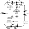
Figure 2.
Figure 2.
Osmoregulatory systems of E. coli. In high osmotic pressure environments, solutes accumulate in E. coli via synthesis (glutamate, trehalose, glycine betaine) or transport from the external medium (e.g., others shown in Fig. 1; Altendorf et al., 2009). K+-H+ symporter Trk and P-type ATPase Kdp mediate K+ uptake. Major facilitator superfamily member ProP, ABC transporter ProU, and betaine-carnitine-choline family members BetT and BetU mediate organic osmolyte uptake. ProP and ProU are similarly broad in substrate specificity, whereas BetT is choline specific (Murdock et al., 2014) and BetU is betaine specific. Mechanosensitive channels, including MscS and MscL, release solutes from the cytoplasm of osmotically downshocked bacteria. Aquaporin AqpZ exacerbates osmotic stress by accelerating transmembrane water flux. BetT and BetU are homologues of BetP from C. glutamicum, whereas ProU is a homologue of OpuA from L. lactis (see Fig. 3).
Figure 3.
Figure 3.
Structures of osmosensory transporters. The structures of BetP from C. glutamicum, ProP from E. coli, and OpuA from L. lactis are illustrated. The protein backbones are colored according to amino acid side-chain polarity unless otherwise indicated: red for acidic residues Asp and Glu; blue for basic residues Arg, Lys, and His; green for polar residues Ser, Thr, Cys, Asn, and Gln; and yellow for nonpolar residues. BetP: a crystal structure of trimeric BetP (Protein Data Bank [PDB] accession no. 2WIT) as viewed from the cytoplasm (A) and of a single BetP subunit as viewed from the membrane (B). In A, the three BetP subunits are colored black, gray, and by amino acid. B shows a single subunit with residues from the N terminus through the end of transmembrane helix II as strands and residues 313–324 as a trace to reveal glycine betaine (space-filling, CPK coloring) within the substrate-binding site. ProP: a homology model of a ProP monomer (PDB accession no. 1Y8S) (C and D, residues 4–236 and 246–452 of the 500-residue ProP protein) and a nuclear magnetic resonance (NMR) structure of the C-terminal domain of ProP (PDB accession no. 1R48) (E, residues 468–497 of the 500-residue ProP protein). ProP is viewed from the membrane with the cytoplasmic surface down (C) and from the cytoplasm (D). The arrow in C marks the position of a substrate analogue in the crystal structure of homologue LacY (PDB accession no. 1PV7). The stars in C and D mark the C-terminal amino acid of the model. (E) The structure of a homodimeric peptide corresponding to residues 468–497 of ProP, determined by NMR spectroscopy (PDB accession no. 1R48). This antiparallel α-helical coiled-coil and transmembrane helix XII contribute to the ProP dimer interface in vivo (Wood, 2011b). OpuA: a schematic representation of transporter OpuA (F) and the structure of periplasmic-binding protein domain OpuAC (G). In F, two cytoplasmic ATP-binding OpuAA subunits, including C-terminal cystathionine-β-synthase (CBS) domains, are blue. Two transmembrane OpuAB domains and the contiguous substrate-binding OpuAC domains are yellow. G shows a crystal structure of domain OpuAC (yellow; PDB accession no. 3L6H) in complex with glycine betaine (spheres). The binding pocket includes three Trp residues (W330, W377, and W484, shown as sticks) that coordinate the trimethylammonium group of glycine betaine.
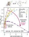
Figure 4.
Figure 4.
Effects of Hofmeister salts on protein unfolding. The effects of low and high concentrations of salts spanning the Hofmeister series on unfolding of the lac repressor DNA binding domain at 40°C. Kobs is the unfolding equilibrium constant in the presence of salt at concentration X, and Kobs,ref is the reference equilibrium constant in low salt buffer. At low salt concentration, all salts exert similar stabilizing effects. These are Coulombic in origin and vary nonlinearly with salt concentration. At high salt concentration, different salts exhibit a wide range of stabilizing (e.g., (NH4)2SO4, KF) to destabilizing (guanidinium HCl (GuHCl)) effects, which are linear in salt concentration and follow the traditional Hofmeister series. The slopes of these high salt linear regions (m-value/RT) correlate with the magnitude and chemical composition of the protein surface that is exposed to the solution in unfolding and the chemical properties of the salt (the places of the cation and anion in the Hofmeister series). Fitted curves allow a separation of Coulombic and Hofmeister effects of these salts. Adapted from Fig. 6 of Record et al. (2013) with permission of The Royal Society of Chemistry (http://dx.doi.org/10.1039/C2FD20128C).

Similar articles

Cited by

References

    1. Altendorf K., Booth I.R., Gralla J.D., Greie J.-C., Rosenthal A.Z., and Wood J.M.. 2009. Osmotic stress. EcoSal Plus. 10.1128/ecosalplus.5.4.5 - DOI - PubMed
    1. Becker M., Maximov S., Becker M., Meyer U., Wittmann A., and Krämer R.. 2014. Analysis of putative protomer crosstalk in the trimeric transporter BetP: The heterotrimer approach. Biochim. Biophys. Acta. 1837:888–898 10.1016/j.bbabio.2014.03.002 - DOI - PubMed
    1. Cayley S., and Record M.T. Jr. 2003. Roles of cytoplasmic osmolytes, water, and crowding in the response of Escherichia coli to osmotic stress: Biophysical basis of osmoprotection by glycine betaine. Biochemistry. 42:12596–12609 10.1021/bi0347297 - DOI - PubMed
    1. Cayley S., and Record M.T. Jr. 2004. Large changes in cytoplasmic biopolymer concentration with osmolality indicate that macromolecular crowding may regulate protein-DNA interactions and growth rate in osmotically stressed Escherichia coli K-12. J. Mol. Recognit. 17:488–496 10.1002/jmr.695 - DOI - PubMed
    1. Culham D.E., Meinecke M., and Wood J.M.. 2012. Impacts of the osmolality and the lumenal ionic strength on osmosensory transporter ProP in proteoliposomes. J. Biol. Chem. 287:27813–27822 10.1074/jbc.M112.387936 - DOI - PMC - PubMed

Publication types

MeSH terms

Substances

LinkOut - more resources