. 2015 May 15;194(10):4698-704.
doi: 10.4049/jimmunol.1400560. Epub 2015 Apr 13.

Recycled IL-7 Can Be Delivered to Neighboring T Cells

Affiliations

Recycled IL-7 Can Be Delivered to Neighboring T Cells

Douglas A Bazdar et al. J Immunol. .

Abstract

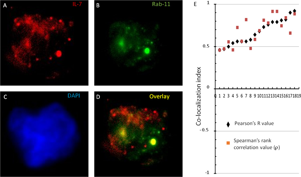
IL-7 is a key homeostatic cytokine that provides signals for T cell survival and proliferation in vivo. In this article, we provide evidence that IL-7 utilization is enhanced by a novel mechanism of cytokine "recycling" during which T cells treated with rIL-7 are rapidly induced to express p-STAT5 and are subsequently able to recycle biologically active cytokine for release to neighboring cells in soluble form. Our observations indicate that the ability of cells to recycle IL-7 is dependent on IL-7R α-chain (CD127) and endocytosis, consistent with a model whereby IL-7 is internalized via receptor interactions before recycling. These observations provide evidence of a novel mechanism that enables cells to optimally use IL-7.


Figures

Figure 1
Figure 1
Evidence of IL-7 transfer between cells. (A) PBMC (2×10⁶/ml) were incubated with rIL-7 (5 ng/ml) for 15 min. Cells were washed 2x and transferred to transwell plates. CFSE-labeled PBMC (responder cells) were mixed with transferred cells in the bottom well at a 1:1 ratio and unlabeled PBMC responder cells were also placed in the top well. P-STAT5 expression was detected by intracellular flow cytometry among CD4+CD3+ cells after 15 min. (B) Summary data of transwell experiments from 5 donors showing the percentages of responder cells that express P-STAT5. Each symbol represents cells from a different donor. (C) PBMC were treated with IL-7 for 15 min. Cells pre-incubated with IL-7 were transferred to CFSE+ responder cells in the presence or absence of an anti-IL-7 neutralizing antibody (10 µg/ml) or an isotype control antibody. (D) Summary data for anti-IL-7 neutralizing antibody studies are shown for cells from 3 different donors. The percent of responder cells expressing P-STAT5 is shown.
Figure 2
Figure 2
Anti-CD127 antibody disrupts the ability of cells to transfer IL-7 to neighboring cells. PBMC from 2 different donors (2×10⁶/ml) were incubated with rIL-7 (5 ng/ml) for 15 min in the presence of anti-CD127 antibody (10 µg/ml) or an isotype control antibody. Cells were washed 2x and transferred to transwell plates. CFSE-labeled PBMC (responder cells) were mixed with transferred cells at a 1:1 ratio and P-STAT5 expression was detected by intracellular flow cytometry among CD4+CD3+ cells after 15 min incubation. Results of cells from each donor are shown in rows (top and bottom).
Figure 3
Figure 3
Inhibitors of endocytosis reduce the capacity of cells to transfer IL-7 to neighboring cells. PBMC were incubated with phenylarsine (PL; 1.3 or 0.13 nM), chlorpromozine (CP; 50 or 75 µM) or DMSO for 15 min and then pulsed with rIL-7 (5 ng/ml) for 15 additional min. These cells were washed and mixed with CFSE+ PBMC in the absence of inhibitors. (A) Representative histograms show P-STAT5 signaling detected by intracellular flow cytometry in responder cells (CFSE+ cells) and in cells used to deliver the IL-7 (CFSE− cells). (B) Summary data of experiments (each symbol represents a different donor) showing inhibition of P-STAT5 induction in CFSE+ cells from inclusion of phenylarsine (PL; left) or chlorpromazine (CP; right) to cells that were pulsed with IL-7. Multi-group comparison (Friedman indicated significant differences with p = 0.002 and 0.0017 for phenylarsine and chlorpromazine experiments, respectively. Student’s t-test for paired data was used to generate p values shown above.
Figure 4
Figure 4
Release of cytokine into supernatants by cells pre-exposed to IL-7. PBMC were incubated with rIL-7 (50 ng/ml) overnight and washed 2x. Cells were then re-plated in complete medium for 5, 30 or 120 min. At these time points the supernatants were centrifuged to remove cells and transferred to responder cells to measure P-STAT5 induction. The cells from these same cultures were washed 2x and transferred to mixed cell cultures with CFSE+ responder cells. P-STAT5 expression was measured in CFSE+ cells by intracellular flow cytometry. (A) P-STAT5 induction in CD4+CFSE+ responder cells was measured after transfer of supernatants (closed symbols and solid lines) or the transfer of cells (1:1 ratio of IL-7-pre-treated cells : CFSE+ responder cells) after 15 min incubation. Statistically significant differences in P-STAT5 induction were observed between the 5 min transfer of cells and the 30 min or 120 min transfer of cells (paired t-test, p = 0.012 and 0.018, respectively). (B) P-STAT5 induction in transferred supernatants from the 30 min incubation time point were treated with anti-IL-7 neutralizing antibody or an isotype control antibody. Control supernatants were from cells that had been incubated overnight in medium alone and then transferred to responder cells in the same manner as the supernatants from IL-7-treated cells.
Figure 5
Figure 5
IL-7 can be presented to neighboring cells even following acid wash. PBMC were incubated overnight with rIL-7 (50 ng/ml). Cells were then washed 2x with acid buffer (pH=3) and 2x with PBS or were washed 4x with PBS. The cells were plated with CFSE+ responder cells at 1:1 ratio and incubated for 15 min or 2 h. P-STAT5 was measured by intracellular flow cytometry. (A) Representative flow cytometry histograms from 1 donor are shown. (B) Summary data from 3 different donors (only 2 donors were tested at the early time point).
Figure 6
Figure 6
Release of biologically active IL-7 by IL-7-treated cells (A) PBMC were incubated with IL-7 (10 ng/ml) for 15 min, washed 2x and re-plated for an additional 15 min or overnight incubation. Supernatants were collected and assessed for IL-7 content by ELISA (p-values derived with Wilcoxon Rank Sum test for non-parametric analyses of paired data, SPSS). (B) PBMC were incubated with rIL-7 (10 ng/ml) in hypertonic sucrose solution for 15 min or isotonic medium. Cells were then washed 2x and re-plated in either hypertonic solution or isotonic medium. Supernatants were collected after 15 min and tested for IL-7 by ELISA (p-values derived from non-parametric paired sample analyses). The p-values shown were derived with Wilcoxon Rank Sum test for non-parametric analyses of paired data, SPSS. Friedman multi-group comparison (C) Four supernatants from (A) were frozen and then later thawed and tested for induction of P-STAT5 in CD4+ T cells after 15 min incubation. The activity in the supernatants (closed symbols) was tested in comparison to a dose-response curve using stock rIL-7 (connected open diamonds). The symbol used in C is matched with the respective symbol in A for each of the 4 supernatants tested.
Figure 7
Figure 7
IL-7 co-localizes with the recycling endosome marker, Rab11, in cytokine pulsed lymphocytes. Cytospin preparations of PBMC were immunostained for IL-7 and Rab11 after 1 hour of incubation with rIL-7 (100 ng/ml) as described in the methods. Representative image of one lymphocyte showing (A) IL-7 (red), (B) Rab11 (green), and (C) DAPI (blue). (D) Merged image indicates evidence of co-localization of fluorescent signals. (E) The correlation coefficients (Pearson’s R value, and Spearman’s rank correlation value(ρ) for pixel co-localization of IL-7 and Rab11 signal in 18 cells is shown and plotted from the cell with the least correlation to the cell with the greatest correlation. Cells that were not pulsed with IL-7 and antibody isotype control stains were used as negative controls and did not demonstrate evidence of staining (not shown).
Figure 8
Figure 8
Glycosylated IL-7 is also recycled for presentation to neighboring cells. PBMC were pulsed with glycosylated IL-7 purified from genetically engineered HEK cells at various concentrations. Cells were then washed and mixed with responder cells as described in Fig 1a. P-STAT5 induction was measured in responding CD4+ T cells. Induction of P-STAT5 in responder cells was significant above the background at each concentration of IL-7 used (p values < 0.05). Each symbol represents cells from a different donor.

Similar articles

Cited by

References

    1. Giliani S, Mori L, de Saint Basile G, Le Deist F, Rodriguez-Perez C, Forino C, Mazzolari E, Dupuis S, Elhasid R, Kessel A, Galambrun C, Gil J, Fischer A, Etzioni A, Notarangelo LD. Interleukin-7 receptor alpha (IL-7Ralpha) deficiency: cellular and molecular bases. Analysis of clinical, immunological, and molecular features in 16 novel patients. Immunol Rev. 2005;203:110–126. - PubMed
    1. Peschon JJ, Morrissey PJ, Grabstein KH, Ramsdell FJ, Maraskovsky E, Gliniak BC, Park LS, Ziegler SF, Williams DE, Ware CB, Meyer JD, Davison BL. Early lymphocyte expansion is severely impaired in interleukin 7 receptor-deficient mice. J Exp Med. 1994;180:1955–1960. - PMC - PubMed
    1. von Freeden-Jeffry U, Vieira P, Lucian LA, McNeil T, Burdach SE, Murray R. Lymphopenia in interleukin (IL)-7 gene-deleted mice identifies IL-7 as a nonredundant cytokine. J Exp Med. 1995;181:1519–1526. - PMC - PubMed
    1. Kondrack RM, Harbertson J, Tan JT, McBreen ME, Surh CD, Bradley LM. Interleukin 7 regulates the survival and generation of memory CD4 cells. J Exp Med. 2003;198:1797–1806. - PMC - PubMed
    1. Tan JT, Dudl E, LeRoy E, Murray R, Sprent J, Weinberg KI, Surh CD. IL-7 is critical for homeostatic proliferation and survival of naive T cells. Proc Natl Acad Sci U S A. 2001;98:8732–8737. - PMC - PubMed

Publication types

LinkOut - more resources